


anti-Emerin antibody


Key features and details
- 产品描述:
- 反应物种:
- 应用:
- 宿主:
- 克隆:
- 同位型:
- 靶点名称:
- 抗原物种:
- 抗原:
-
Brand:

 Product Details
| 产品描述 | Rabbit Polyclonal antibody recognizes Emerin |
|---|---|
| 反应物种 | Hu, Ms, Rat |
| 应用 | FACS, ICC/IF, IHC-P, WB |
| 宿主 | Rabbit |
| 克隆 | Polyclonal |
| 同位型 | IgG |
| 靶点名称 | Emerin |
| 抗原物种 | Human |
| 抗原 | Synthetic peptide corresponding to aa. 1-48 of Human Emerin. (MDNYADLSDTELTTLLRRYNIPHGPVVGSTRRLYEKKIFEYETQRRRL) |
| 偶联标记 | Un-conjugated |
| 別名 | Emerin; LEMD5; EDMD; STA |
| 应用建议 |
| ||||||||||
|---|---|---|---|---|---|---|---|---|---|---|---|
| 应用说明 | IHC-P: Antigen Retrieval: Heat mediation was performed in Citrate buffer (pH 6.0) for 20 min, or performed in EDTA buffer (pH 8.0). * The dilutions indicate recommended starting dilutions and the optimal dilutions or concentrations should be determined by the scientist. |
| 形式 | Liquid |
|---|---|
| 纯化 | Affinity purification with immunogen. |
| 缓冲液 | 0.9% NaCl, 0.2% Na2HPO4, 0.05% Sodium azide and 5% BSA. |
| 抗菌剂 | 0.05% Sodium azide |
| 稳定剂 | 5% BSA |
| 浓度 | 0.5 mg/ml |
| 存放说明 | For continuous use, store undiluted antibody at 2-8°C for up to a week. For long-term storage, aliquot and store at -20°C or below. Storage in frost free freezers is not recommended. Avoid repeated freeze/thaw cycles. Suggest spin the vial prior to opening. The antibody solution should be gently mixed before use. |
| 注意事项 | For laboratory research only, not for drug, diagnostic or other use. |
| 数据库连接 | |
|---|---|
| 基因名称 | EMD |
| 全名 | emerin |
| 背景介绍 | Emerin is a serine-rich nuclear membrane protein and a member of the nuclear lamina-associated protein family. It mediates membrane anchorage to the cytoskeleton. Dreifuss-Emery muscular dystrophy is an X-linked inherited degenerative myopathy resulting from mutation in the emerin gene. [provided by RefSeq, Jul 2008] |
| 生物功能 | Stabilizes and promotes the formation of a nuclear actin cortical network. Stimulates actin polymerization in vitro by binding and stabilizing the pointed end of growing filaments. Inhibits beta-catenin activity by preventing its accumulation in the nucleus. Acts by influencing the nuclear accumulation of beta-catenin through a CRM1-dependent export pathway. Links centrosomes to the nuclear envelope via a microtubule association. EMD and BAF are cooperative cofactors of HIV-1 infection. Association of EMD with the viral DNA requires the presence of BAF and viral integrase. The association of viral DNA with chromatin requires the presence of BAF and EMD. Required for proper localization of non-farnesylated prelamin-A/C. [UniProt] |
| 细胞定位 | Nucleus inner membrane; Single-pass membrane protein; Nucleoplasmic side. Nucleus outer membrane. Colocalized with BANF1 at the central region of the assembling nuclear rim, near spindle-attachment sites. The accumulation of different intermediates of prelamin-A/C (non- farnesylated or carboxymethylated farnesylated prelamin-A/C) in fibroblasts modify its localization in the nucleus. [UniProt] |
| 预测分子量 | 29 kDa |
| 翻译后修饰 | Found in four different phosphorylated forms, three of which appear to be associated with the cell cycle. [UniProt] |

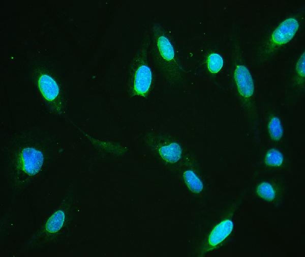
ARG58572 anti-Emerin antibody ICC/IF image
Immunofluorescence: U2OS cells were blocked with 10% goat serum and then stained with ARG58572 anti-Emerin antibody (green) at 2 µg/ml dilution, overnight at 4°C. DAPI (blue) for nuclear staining.
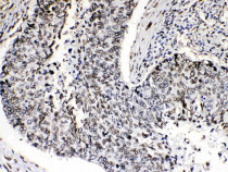
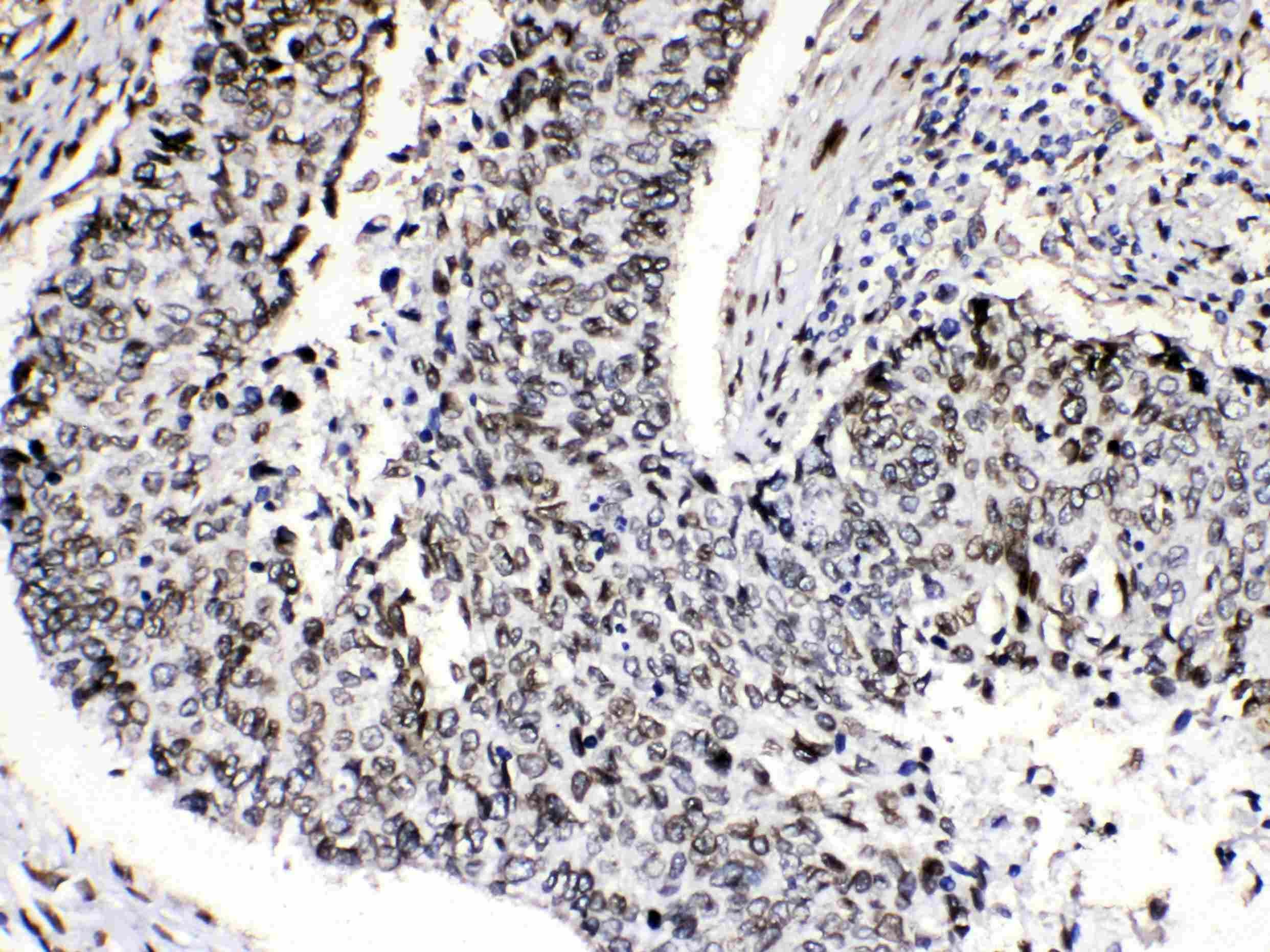
ARG58572 anti-Emerin antibody IHC-P image
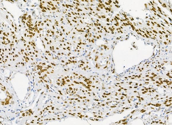
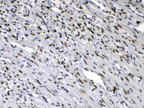
Immunohistochemistry: Paraffin-embedded Human intetsinal cancer tissuesstained with ARG58572 anti-Emerin antibody at 1 µg/ml dilution.

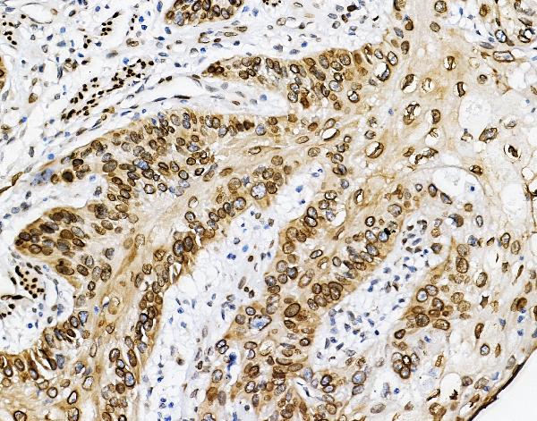
ARG58572 anti-Emerin antibody IHC-P image
Immunohistochemistry: Paraffin-embedded Human endometrial carcinoma tissue. Antigen Retrieval: Heat mediation was performed in Citrate buffer (pH 6.0) for 20 min. The tissue section was blocked with 10% goat serum. The tissue section was then stained with ARG58572 anti-Emerin antibody at 2 µg/ml dilution, overnight at 4°C.
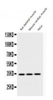
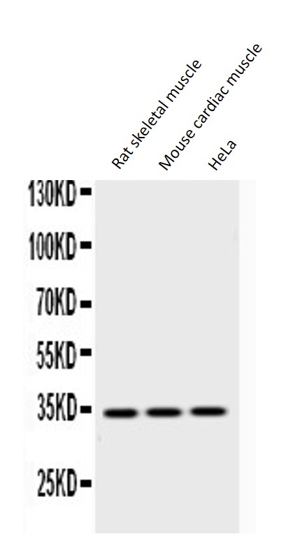
ARG58572 anti-Emerin antibody WB image
Western blot: Rat skeletal muscle extract, Mouse cardiac muscle extract and HeLa whole cell lysates stained with ARG58572 anti-Emerin antibody at 0.5 µg/ml dilution.
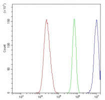
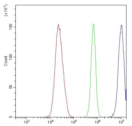
ARG58572 anti-Emerin antibody FACS image
Flow Cytometry: U2OS cells were blocked with 10% normal goat serum and then stained with ARG58572 anti-Emerin antibody (blue) at 1 µg/10^6 cells for 30 min at 20°C, followed by incubation with DyLight®488 labelled secondary antibody. Isotype control antibody (green) was rabbit IgG (1 µg/10^6 cells) used under the same conditions. Unlabelled sample (red) was also used as a control.
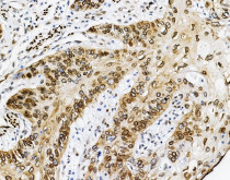
ARG58572 anti-Emerin antibody IHC-P image
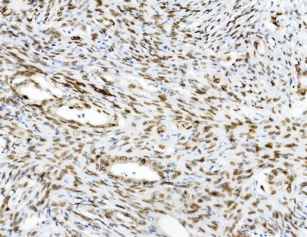
Immunohistochemistry: Paraffin-embedded Human lung cancer tissues stained with ARG58572 anti-Emerin antibody at 1 µg/ml dilution.
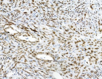
ARG58572 anti-Emerin antibody IHC-P image
Immunohistochemistry: Paraffin-embedded Human colon cancer tissue. Antigen Retrieval: Heat mediation was performed in Citrate buffer (pH 6.0) for 20 min. The tissue section was blocked with 10% goat serum. The tissue section was then stained with ARG58572 anti-Emerin antibody at 1 µg/ml dilution, overnight at 4°C.
ARG58572 anti-Emerin antibody IHC-P image
Immunohistochemistry: Paraffin-embedded Human oesophagus squama cancer tissue. Antigen Retrieval: Heat mediation was performed in Citrate buffer (pH 6.0) for 20 min. The tissue section was blocked with 10% goat serum. The tissue section was then stained with ARG58572 anti-Emerin antibody at 1 µg/ml dilution, overnight at 4°C.

ARG58572 anti-Emerin antibody IHC-P image
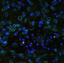
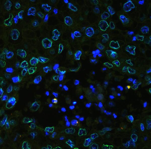
Immunohistochemistry: Paraffin-embedded Human oesophagus squama cancer tissue. Antigen Retrieval: Heat mediation was performed in Citrate buffer (pH 6.0) for 20 min. The tissue section was blocked with 10% goat serum. The tissue section was then stained with ARG58572 anti-Emerin antibody (green) at 1 µg/ml dilution, overnight at 4°C. The section was counterstained with DAPI (blue).
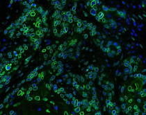
ARG58572 anti-Emerin antibody IHC-P image
Immunohistochemistry: Paraffin-embedded Human oesophagus squama cancer tissue. Antigen Retrieval: Heat mediation was performed in Citrate buffer (pH 6.0) for 20 min. The tissue section was blocked with 10% goat serum. The tissue section was then stained with ARG58572 anti-Emerin antibody (green) at 2 µg/ml dilution, overnight at 4°C. The section was counterstained with DAPI (blue).
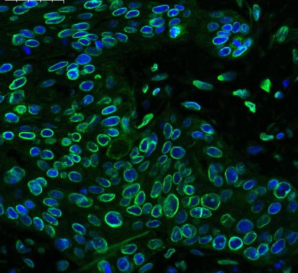
ARG58572 anti-Emerin antibody IHC-P image
Immunohistochemistry: Paraffin-embedded Human lung cancer tissue. Antigen Retrieval: Heat mediation was performed in EDTA buffer (pH 8.0). The tissue section was blocked with 10% goat serum. The tissue section was then stained with ARG58572 anti-Emerin antibody (green) at 2 µg/ml dilution, overnight at 4°C. The section was counterstained with DAPI (blue).


 New Products


