


anti-ERK1 antibody [SQab1866]
![anti-ERK1 antibody [SQab1866]](/upload/image/products/ARG66318_WB_5L_210_205.jpg)
![anti-ERK1 antibody [SQab1866]](/upload/image/products/ARG66318_IF_HeLa.jpg)
![anti-ERK1 antibody [SQab1866]](/upload/image/products/ARG66318_FACS_HeLa.jpg)
![anti-ERK1 antibody [SQab1866]](/upload/image/products/ARG66318_IHC-P_CervixCancer.jpg)
![anti-ERK1 antibody [SQab1866]](/upload/image/products/ARG66318_WB_7L.jpg)
![anti-ERK1 antibody [SQab1866]](/upload/image/products/ARG66318_IP_A375.jpg)
![anti-ERK1 antibody [SQab1866]](/upload/image/products/ARG66318_WB_5L.jpg)
Key features and details
- 产品描述:
- 反应物种:
- 应用:
- 宿主:
- 克隆:
- 克隆号:
- 同位型:
- 靶点名称:
- 抗原物种:
-
Brand:

 Product Details
| 产品描述 | Recombinant Rabbit Monoclonal antibody [SQab1866] recognizes ERK1 |
|---|---|
| 反应物种 | Hu, Ms, Rat |
| 应用 | FACS, ICC/IF, IHC-P, IP, WB |
| 宿主 | Rabbit |
| 克隆 | Monoclonal |
| 克隆号 | SQab1866 |
| 同位型 | IgG |
| 靶点名称 | ERK1 |
| 抗原物种 | Human |
| 抗原 | Synthetic peptide around the N-terminus of ERK1. |
| 偶联标记 | Un-conjugated |
| 別名 | MAPK 3; ERK1; P44MAPK; Microtubule-associated protein 2 kinase; Insulin-stimulated MAP2 kinase; HUMKER1A; PRKM3; P44ERK1; EC 2.7.11.24; p44-MAPK; Extracellular signal-regulated kinase 1; p44-ERK1; HS44KDAP; MAP kinase isoform p44; Mitogen-activated protein kinase 3; ERT2; MAP kinase 3; ERK-1 |
| 应用建议 |
| ||||||||||||
|---|---|---|---|---|---|---|---|---|---|---|---|---|---|
| 应用说明 | IHC-P: Antigen Retrieval: Heat mediated was performed using Tris/EDTA buffer pH 9.0. * The dilutions indicate recommended starting dilutions and the optimal dilutions or concentrations should be determined by the scientist. |
| 形式 | Liquid |
|---|---|
| 纯化 | Purification with Protein A. |
| 缓冲液 | PBS, 0.01% Sodium azide, 40% Glycerol and 0.05% BSA. |
| 抗菌剂 | 0.01% Sodium azide |
| 稳定剂 | 40% Glycerol and 0.05% BSA |
| 存放说明 | For continuous use, store undiluted antibody at 2-8°C for up to a week. For long-term storage, aliquot and store at -20°C. Storage in frost free freezers is not recommended. Avoid repeated freeze/thaw cycles. Suggest spin the vial prior to opening. The antibody solution should be gently mixed before use. |
| 注意事项 | For laboratory research only, not for drug, diagnostic or other use. |
| 数据库连接 | |
|---|---|
| 基因名称 | MAPK3 |
| 全名 | mitogen-activated protein kinase 3 |
| 背景介绍 | ERK1 is a member of the MAP kinase family. MAP kinases, also known as extracellular signal-regulated kinases (ERKs), act in a signaling cascade that regulates various cellular processes such as proliferation, differentiation, and cell cycle progression in response to a variety of extracellular signals. This kinase is activated by upstream kinases, resulting in its translocation to the nucleus where it phosphorylates nuclear targets. Alternatively spliced transcript variants encoding different protein isoforms have been described. [provided by RefSeq, Jul 2008] |
| 生物功能 | Serine/threonine kinase which acts as an essential component of the MAP kinase signal transduction pathway. MAPK1/ERK2 and MAPK3/ERK1 are the 2 MAPKs which play an important role in the MAPK/ERK cascade. They participate also in a signaling cascade initiated by activated KIT and KITLG/SCF. Depending on the cellular context, the MAPK/ERK cascade mediates diverse biological functions such as cell growth, adhesion, survival and differentiation through the regulation of transcription, translation, cytoskeletal rearrangements. The MAPK/ERK cascade plays also a role in initiation and regulation of meiosis, mitosis, and postmitotic functions in differentiated cells by phosphorylating a number of transcription factors. About 160 substrates have already been discovered for ERKs. Many of these substrates are localized in the nucleus, and seem to participate in the regulation of transcription upon stimulation. However, other substrates are found in the cytosol as well as in other cellular organelles, and those are responsible for processes such as translation, mitosis and apoptosis. Moreover, the MAPK/ERK cascade is also involved in the regulation of the endosomal dynamics, including lysosome processing and endosome cycling through the perinuclear recycling compartment (PNRC); as well as in the fragmentation of the Golgi apparatus during mitosis. The substrates include transcription factors (such as ATF2, BCL6, ELK1, ERF, FOS, HSF4 or SPZ1), cytoskeletal elements (such as CANX, CTTN, GJA1, MAP2, MAPT, PXN, SORBS3 or STMN1), regulators of apoptosis (such as BAD, BTG2, CASP9, DAPK1, IER3, MCL1 or PPARG), regulators of translation (such as EIF4EBP1) and a variety of other signaling-related molecules (like ARHGEF2, FRS2 or GRB10). Protein kinases (such as RAF1, RPS6KA1/RSK1, RPS6KA3/RSK2, RPS6KA2/RSK3, RPS6KA6/RSK4, SYK, MKNK1/MNK1, MKNK2/MNK2, RPS6KA5/MSK1, RPS6KA4/MSK2, MAPKAPK3 or MAPKAPK5) and phosphatases (such as DUSP1, DUSP4, DUSP6 or DUSP16) are other substrates which enable the propagation the MAPK/ERK signal to additional cytosolic and nuclear targets, thereby extending the specificity of the cascade. [UniProt] |
| 预测分子量 | 43 kDa |
| 翻译后修饰 | Phosphorylated upon KIT and FLT3 signaling (By similarity). Dually phosphorylated on Thr-202 and Tyr-204, which activates the enzyme. Ligand-activated ALK induces tyrosine phosphorylation. Dephosphorylated by PTPRJ at Tyr-204. [UniProt] |
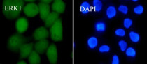
ARG66318 anti-ERK1 antibody [SQab1866] ICC/IF image
Immunofluorescence: HeLa cells were fixed with 4% paraformaldehyde for 30 min at RT, permeabilized with 0.1% Triton X-100 for 10 min at RT then blocked with 10% goat serum for 30 min at RT. Cells were stained with ARG66318 anti-ERK1 antibody [SQab1866] (green) at 1:50 and 4°C. DAPI (blue) was used as the nuclear counter stain.
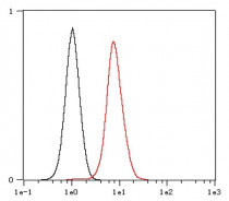
ARG66318 anti-ERK1 antibody [SQab1866] FACS image
Flow Cytometry: HeLa cells were fixed with 4% paraformaldehyde (10 min) and then permeabilized with 0.1% TritonX-100 for 15 min. The cells were stained with ARG66318 anti-ERK1 antibody [SQab1866] (red) at 1:1,000 dilution in 1x PBS/1% BSA for 30 min at RT, followed by Alexa Fluor® 488 labelled secondary antibody. Unlabelled sample (black) was used as a control.
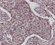
ARG66318 anti-ERK1 antibody [SQab1866] IHC-P image
Immunohistochemistry: Formalin-fixed and paraffin-embedded Cervix cancer tissue stained with ARG66318 anti-ERK1 antibody [SQab1866] at 1:200 dilution. Antigen Retrieval: Heat mediated was performed using Tris/EDTA buffer pH 9.0.
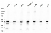
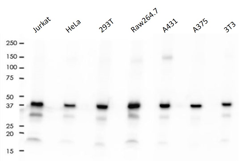
ARG66318 anti-ERK1 antibody [SQab1866] WB image
Western blot: 10 µg of Jurkat, HeLa, 293T, Raw264.7, A431, A375 and 3T3 cell lysates stained with ARG66318 anti-ERK1 antibody [SQab1866] at 1:2000 dilution.
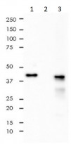
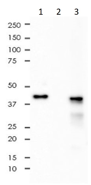
ARG66318 anti-ERK1 antibody [SQab1866] IP image
Immunoprecipitation: 0.4 mg of A375 whole cell lysate immunoprecipitated (1:50) and stained with ARG66318 anti-ERK1 antibody [SQab1866]. 1) ARG66318 IP in A375 whole cell lysate. 2) PBS instead of ARG66318 in A375 whole cell lysate 3) A375 whole cell lysate, 10 µg (input).
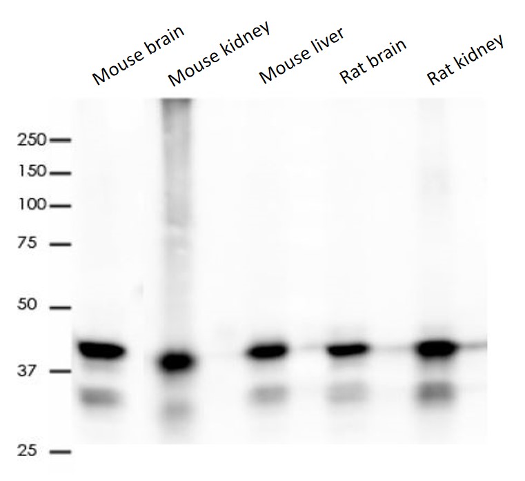
ARG66318 anti-ERK1 antibody [SQab1866] WB image
Western blot: 10 µg of Mouse brain, Mouse kidney, Mouse liver, Rat brain and Rat kidney lysates stained with ARG66318 anti-ERK1 antibody [SQab1866] at 1:2000 dilution.


 New Products


