


anti-EWSR1 / EWS antibody


Key features and details
- 产品描述:
- 反应物种:
- 应用:
- 宿主:
- 克隆:
- 同位型:
- 靶点名称:
- 抗原物种:
- 抗原:
-
Brand:

 Product Details
| 产品描述 | Rabbit Polyclonal antibody recognizes EWSR1 / EWS |
|---|---|
| 反应物种 | Hu, Ms, Rat |
| 应用 | FACS, ICC/IF, IHC-Fr, IHC-P, WB |
| 宿主 | Rabbit |
| 克隆 | Polyclonal |
| 同位型 | IgG |
| 靶点名称 | EWSR1 / EWS |
| 抗原物种 | Human |
| 抗原 | Synthetic peptide corresponding to aa. 369-399 of Human EWSR1 / EWS. (NDSVTLDDLADFFKQCGVVKMNKRTGQPMIH) |
| 偶联标记 | Un-conjugated |
| 別名 | RNA-binding protein EWS; bK984G1.4; EWS-FLI1; Ewing sarcoma breakpoint region 1 protein; EWS; EWS oncogene |
| 应用建议 |
| ||||||||||||
|---|---|---|---|---|---|---|---|---|---|---|---|---|---|
| 应用说明 | IHC-P: Antigen Retrieval: Heat mediation was performed in Citrate buffer (pH 6.0) for 20 min. * The dilutions indicate recommended starting dilutions and the optimal dilutions or concentrations should be determined by the scientist. | ||||||||||||
| 实际分子量 | ~ 93 kDa |
| 形式 | Liquid |
|---|---|
| 纯化 | Affinity purification with immunogen. |
| 缓冲液 | 0.2% Na2HPO4, 0.9% NaCl, 0.05% Sodium azide and 5% BSA. |
| 抗菌剂 | 0.05% Sodium azide |
| 稳定剂 | 5% BSA |
| 浓度 | 0.5 mg/ml |
| 存放说明 | For continuous use, store undiluted antibody at 2-8°C for up to a week. For long-term storage, aliquot and store at -20°C or below. Storage in frost free freezers is not recommended. Avoid repeated freeze/thaw cycles. Suggest spin the vial prior to opening. The antibody solution should be gently mixed before use. |
| 注意事项 | For laboratory research only, not for drug, diagnostic or other use. |
| 数据库连接 | |
|---|---|
| 基因名称 | EWSR1 |
| 全名 | EWS RNA-binding protein 1 |
| 背景介绍 | This gene encodes a multifunctional protein that is involved in various cellular processes, including gene expression, cell signaling, and RNA processing and transport. The protein includes an N-terminal transcriptional activation domain and a C-terminal RNA-binding domain. Chromosomal translocations between this gene and various genes encoding transcription factors result in the production of chimeric proteins that are involved in tumorigenesis. These chimeric proteins usually consist of the N-terminal transcriptional activation domain of this protein fused to the C-terminal DNA-binding domain of the transcription factor protein. Mutations in this gene, specifically a t(11;22)(q24;q12) translocation, are known to cause Ewing sarcoma as well as neuroectodermal and various other tumors. Alternative splicing of this gene results in multiple transcript variants. Related pseudogenes have been identified on chromosomes 1 and 14. [provided by RefSeq, Jul 2009] |
| 生物功能 | Might normally function as a transcriptional repressor. EWS-fusion-proteins (EFPS) may play a role in the tumorigenic process. They may disturb gene expression by mimicking, or interfering with the normal function of CTD-POLII within the transcription initiation complex. They may also contribute to an aberrant activation of the fusion protein target genes. [UniProt] |
| 细胞定位 | Nucleus. Cytoplasm. Cell membrane. Note=Relocates from cytoplasm to ribosomes upon PTK2B/FAK2 activation. [UniProt] |
| 预测分子量 | 68 kDa |
| 翻译后修饰 | Phosphorylated; calmodulin-binding inhibits phosphorylation of Ser-266. Highly methylated on arginine residues. Methylation is mediated by PRMT1 and, at lower level by PRMT8. [UniProt] |

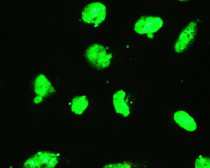
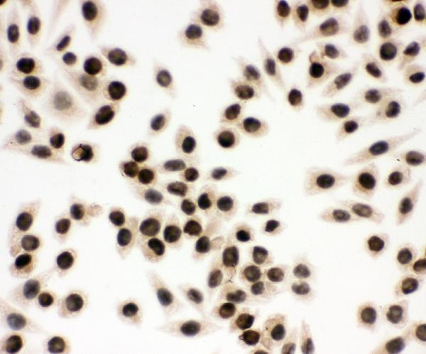
ARG43229 anti-EWSR1 / EWS antibody ICC image
Immunocytochemistry: SMMC-7721 cells stained with ARG43229 anti-EWSR1 / EWS antibody at 1 µg/ml dilution, overnight at 4°C.
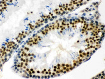
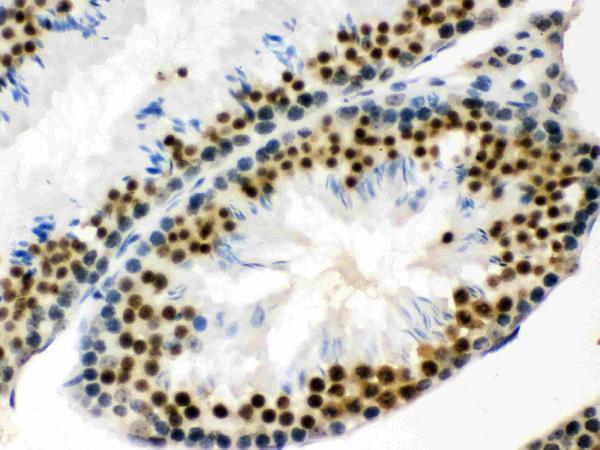
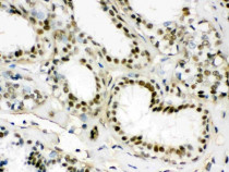
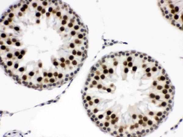
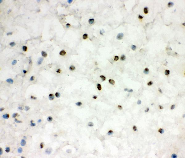
ARG43229 anti-EWSR1 / EWS antibody IHC-P image
Immunohistochemistry: Paraffin-embedded Mouse testis tissue. Antigen Retrieval: Heat mediation was performed in Citrate buffer (pH 6.0) for 20 min. The tissue section was blocked with 10% goat serum. The tissue section was then stained with ARG43229 anti-EWSR1 / EWS antibody at 1 µg/ml dilution, overnight at 4°C.

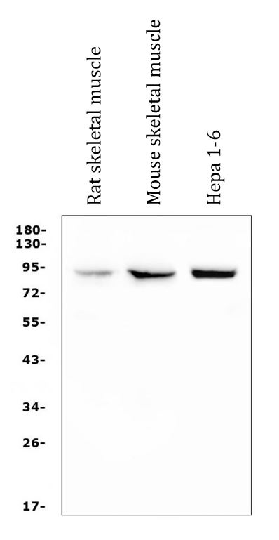
ARG43229 anti-EWSR1 / EWS antibody WB image
Western blot: 50 µg of sample under reducing conditions. Rat skeletal muscle, Mouse skeletal muscle and Hepa 1-6 whole cell lysate stained with ARG43229 anti-EWSR1 / EWS antibody at 0.5 µg/ml dilution, overnight at 4°C.
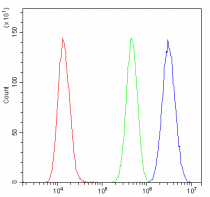
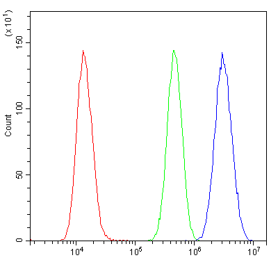
ARG43229 anti-EWSR1 / EWS antibody FACS image
Flow Cytometry: U2OS cells were blocked with 10% normal goat serum and then stained with ARG43229 anti-EWSR1 / EWS antibody (blue) at 1 µg/10^6 cells for 30 min at 20°C, followed by incubation with DyLight®488 labelled secondary antibody. Isotype control antibody (green) was rabbit IgG (1 µg/10^6 cells) used under the same conditions. Unlabelled sample (red) was also used as a control.
ARG43229 anti-EWSR1 / EWS antibody ICC/IF image
Immunofluorescence: U2OS cells stained with ARG43229 anti-EWSR1 / EWS antibody (green) at 2 µg/ml dilution, overnight at 4°C.

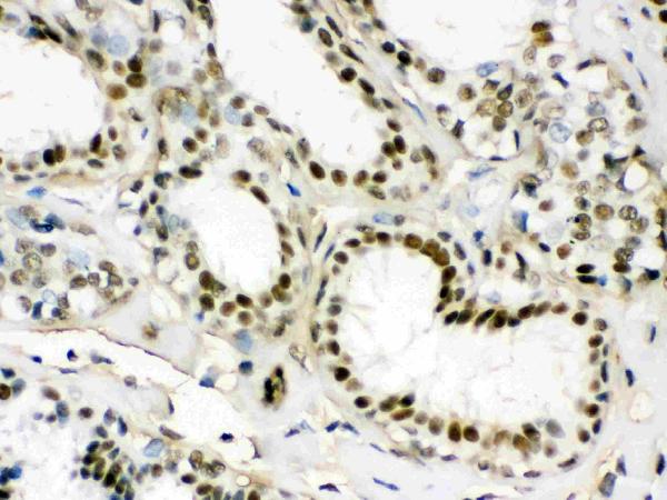
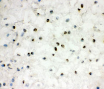
ARG43229 anti-EWSR1 / EWS antibody IHC-P image
Immunohistochemistry: Paraffin-embedded Rat testis tissue. Antigen Retrieval: Heat mediation was performed in Citrate buffer (pH 6.0) for 20 min. The tissue section was blocked with 10% goat serum. The tissue section was then stained with ARG43229 anti-EWSR1 / EWS antibody at 1 µg/ml dilution, overnight at 4°C.
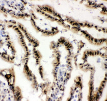
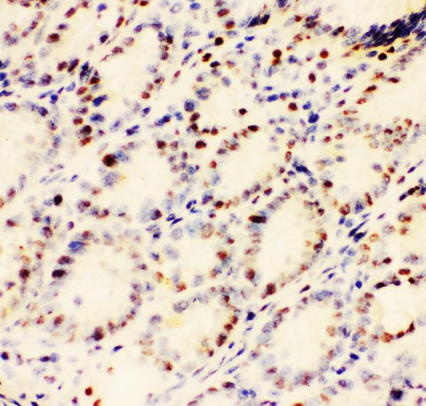
ARG43229 anti-EWSR1 / EWS antibody IHC-P image
Immunohistochemistry: Paraffin-embedded Human mammary cancer tissue. Antigen Retrieval: Heat mediation was performed in Citrate buffer (pH 6.0) for 20 min. The tissue section was blocked with 10% goat serum. The tissue section was then stained with ARG43229 anti-EWSR1 / EWS antibody at 1 µg/ml dilution, overnight at 4°C.

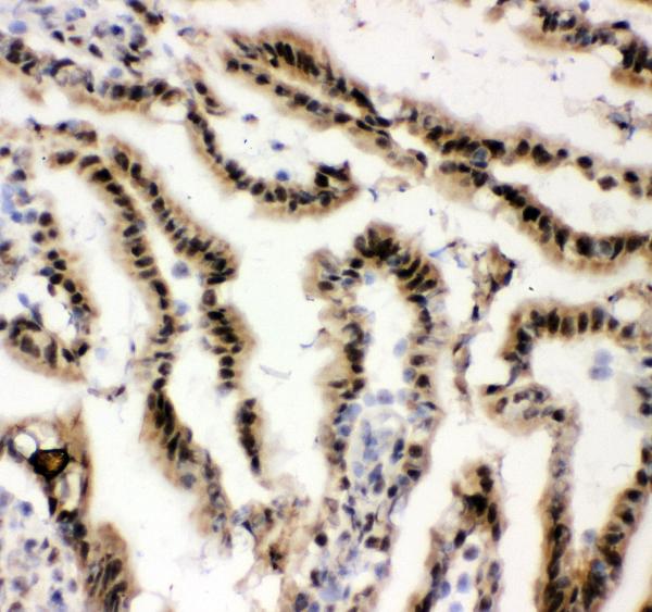
ARG43229 anti-EWSR1 / EWS antibody IHC-Fr image
Immunohistochemistry: Frozen section of Mouse intestine tissue. The tissue section was blocked with 10% goat serum. The tissue section was then stained with ARG43229 anti-EWSR1 / EWS antibody at 1 µg/ml dilution, overnight at 4°C.
ARG43229 anti-EWSR1 / EWS antibody IHC-Fr image
Immunohistochemistry: Frozen section of Rat intestine tissue. The tissue section was blocked with 10% goat serum. The tissue section was then stained with ARG43229 anti-EWSR1 / EWS antibody at 1 µg/ml dilution, overnight at 4°C.
ARG43229 anti-EWSR1 / EWS antibody IHC-Fr image
Immunohistochemistry: Frozen section of Human placenta tissue. The tissue section was blocked with 10% goat serum. The tissue section was then stained with ARG43229 anti-EWSR1 / EWS antibody at 1 µg/ml dilution, overnight at 4°C.
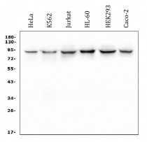
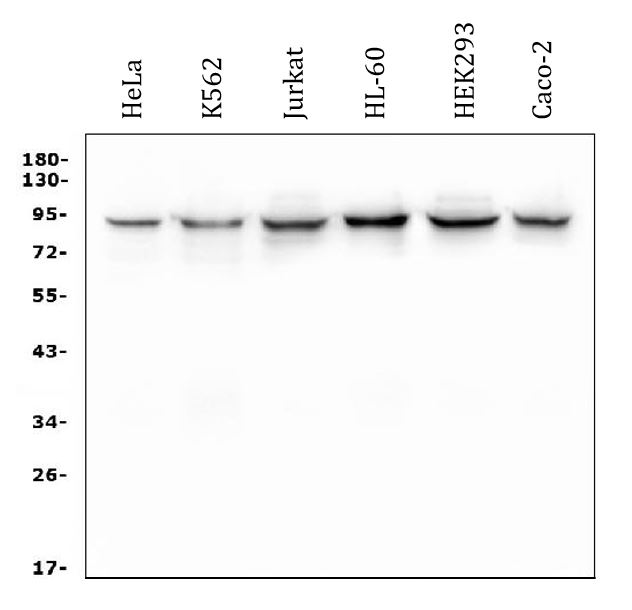
ARG43229 anti-EWSR1 / EWS antibody WB image
Western blot: 50 µg of sample under reducing conditions. HeLa, K562, Jurkat, HL-60, HEK293 and Caco-2 whole cell lysate stained with ARG43229 anti-EWSR1 / EWS antibody at 0.5 µg/ml dilution, overnight at 4°C.


 New Products


