


anti-Fibronectin antibody


Key features and details
- 产品描述:
- 反应物种:
- 应用:
- 宿主:
- 克隆:
- 同位型:
- 靶点名称:
- 抗原物种:
- 抗原:
-
Brand:

 Product Details
| 产品描述 | Mouse Monoclonal antibody recognizes Fibronectin |
|---|---|
| 反应物种 | Hu, Ms, Rat |
| 应用 | IHC-P, WB |
| 宿主 | Mouse |
| 克隆 | Monoclonal |
| 同位型 | IgG |
| 靶点名称 | Fibronectin |
| 抗原物种 | Human |
| 抗原 | Synthetic peptide from Human Fibronectin |
| 偶联标记 | Un-conjugated |
| 別名 | ED-B; CIG; GFND; Cold-insoluble globulin; FNZ; LETS; GFND2; Fibronectin; MSF; FINC; FN |
| 应用建议 |
| ||||||
|---|---|---|---|---|---|---|---|
| 应用说明 | IHC-P: Antigen Retrieval: Boil tissue section in Sodium citrate buffer (pH 6.0) for 20 min. * The dilutions indicate recommended starting dilutions and the optimal dilutions or concentrations should be determined by the scientist. |
| 形式 | Liquid |
|---|---|
| 纯化 | Affinity purification with immunogen. |
| 缓冲液 | PBS (pH 7.4), 0.02% Sodium azide and 50% Glycerol. |
| 抗菌剂 | 0.02% Sodium azide |
| 稳定剂 | 50% Glycerol |
| 浓度 | 1 mg/ml |
| 存放说明 | For continuous use, store undiluted antibody at 2-8°C for up to a week. For long-term storage, aliquot and store at -20°C or below. Storage in frost free freezers is not recommended. Avoid repeated freeze/thaw cycles. Suggest spin the vial prior to opening. The antibody solution should be gently mixed before use. |
| 注意事项 | For laboratory research only, not for drug, diagnostic or other use. |
| 数据库连接 | |
|---|---|
| 基因名称 | FN1 |
| 全名 | fibronectin 1 |
| 背景介绍 | This gene encodes fibronectin, a glycoprotein present in a soluble dimeric form in plasma, and in a dimeric or multimeric form at the cell surface and in extracellular matrix. Fibronectin is involved in cell adhesion and migration processes including embryogenesis, wound healing, blood coagulation, host defense, and metastasis. The gene has three regions subject to alternative splicing, with the potential to produce 20 different transcript variants. However, the full-length nature of some variants has not been determined. [provided by RefSeq, Jul 2008] |
| 生物功能 | Fibronectins bind cell surfaces and various compounds including collagen, fibrin, heparin, DNA, and actin. Fibronectins are involved in cell adhesion, cell motility, opsonization, wound healing, and maintenance of cell shape. Involved in osteoblast compaction through the fibronectin fibrillogenesis cell-mediated matrix assembly process, essential for osteoblast mineralization. Participates in the regulation of type I collagen deposition by osteoblasts. Anastellin binds fibronectin and induces fibril formation. This fibronectin polymer, named superfibronectin, exhibits enhanced adhesive properties. Both anastellin and superfibronectin inhibit tumor growth, angiogenesis and metastasis. Anastellin activates p38 MAPK and inhibits lysophospholipid signaling. [UniProt] |
| 产品亮点 | Related Antibody Duos and Panels: ARG30346 Myofibroblast / Fibrosis Antibody Panel Related products: Fibronectin antibodies; Fibronectin ELISA Kits; Fibronectin Duos / Panels; Anti-Mouse IgG secondary antibodies; Related news: New antibody panels for Myofibroblasts and CAFs |
| 预测分子量 | 272 kDa |
| 翻译后修饰 | Sulfated. It is not known whether both or only one of Thr-2064 and Thr-2065 are/is glycosylated. Forms covalent cross-links mediated by a transglutaminase, such as F13A or TGM2, between a glutamine and the epsilon-amino group of a lysine residue, forming homopolymers and heteropolymers (e.g. fibrinogen-fibronectin, collagen-fibronectin heteropolymers). Phosphorylated by FAM20C in the extracellular medium. Proteolytic processing produces the C-terminal NC1 peptide, anastellin. Some lysine residues are oxidized to allysine by LOXL3, promoting fibronectin activation and matrix formation. |

ARG66162 anti-Fibronectin antibody WB image
Western blot: 20 µg of Huh7 and Huh7 shHMGB1 cell lysates stained with ARG66162 anti-Fibronectin antibody (1:1000), ARG55914 anti-E-cadherin antibody (1:1000) and ARG65636 anti-HMGB1 antibody (1:2000).

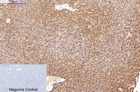
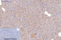
ARG66162 anti-Fibronectin antibody IHC-P image
Immunohistochemistry: Paraffin-embedded Rat liver tissue stained with ARG66162 anti-Fibronectin antibody at 1:200 (4°C, overnight). Antigen Retrieval: Boil tissue section in Sodium citrate buffer (pH 6.0) for 20 min. Secondary antibody was diluted at 1:200 (RT, 30min). Negative control: Secondary antibody only.
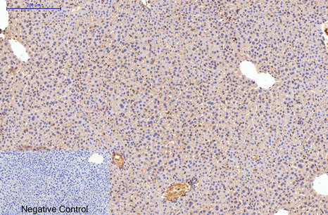
ARG66162 anti-Fibronectin antibody IHC-P image
Immunohistochemistry: Paraffin-embedded Mouse liver tissue stained with ARG66162 anti-Fibronectin antibody at 1:200 (4°C, overnight). Antigen Retrieval: Boil tissue section in Sodium citrate buffer (pH 6.0) for 20 min. Secondary antibody was diluted at 1:200 (RT, 30min). Negative control: Secondary antibody only.
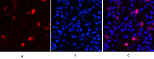
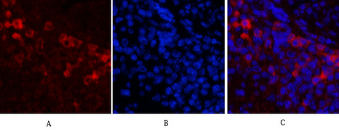
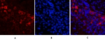
ARG66162 anti-Fibronectin antibody IHC image
Immunohistochemistry: Human appendix tissue stained with ARG66162 anti-Fibronectin antibody (red) at 1:200 (4°C, overnight). Picture A: Target. Picture B: DAPI. Picture C: merge of A and B.

ARG66162 anti-Fibronectin antibody IHC image
Immunohistochemistry: Human appendix tissue stained with ARG66162 anti-Fibronectin antibody (red) at 1:200 (4°C, overnight). Picture A: Target. Picture B: DAPI. Picture C: merge of A and B.
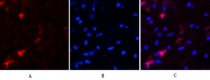
ARG66162 anti-Fibronectin antibody IHC image
Immunohistochemistry: Human appendix tissue stained with ARG66162 anti-Fibronectin antibody (red) at 1:200 (4°C, overnight). Picture A: Target. Picture B: DAPI. Picture C: merge of A and B.

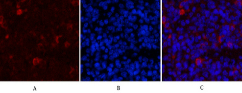
ARG66162 anti-Fibronectin antibody IHC image
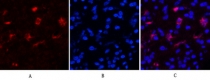
Immunohistochemistry: Mouse spleen tissue stained with ARG66162 anti-Fibronectin antibody (red) at 1:200 (4°C, overnight). Picture A: Target. Picture B: DAPI. Picture C: merge of A and B.

ARG66162 anti-Fibronectin antibody IHC image
Immunohistochemistry: Mouse spleen tissue stained with ARG66162 anti-Fibronectin antibody (red) at 1:200 (4°C, overnight). Picture A: Target. Picture B: DAPI. Picture C: merge of A and B.
ARG66162 anti-Fibronectin antibody IHC image
Immunohistochemistry: Mouse spleen tissue stained with ARG66162 anti-Fibronectin antibody (red) at 1:200 (4°C, overnight). Picture A: Target. Picture B: DAPI. Picture C: merge of A and B.


Anticancer Effects and Molecular Mechanisms of Apigenin in Cervical Cancer Cells
WB, IHC-P / Human

 New Products


