


anti-FLCN antibody


Key features and details
- 产品描述:
- 反应物种:
- 应用:
- 宿主:
- 克隆:
- 同位型:
- 靶点名称:
- 抗原物种:
- 抗原:
-
Brand:

 Product Details
| 产品描述 | Rabbit Polyclonal antibody recognizes FLCN |
|---|---|
| 反应物种 | Hu, Ms, Rat |
| 应用 | FACS, ICC/IF, IHC-P, WB |
| 宿主 | Rabbit |
| 克隆 | Polyclonal |
| 同位型 | IgG |
| 靶点名称 | FLCN |
| 抗原物种 | Human |
| 抗原 | Recombinant protein corresponding to C8-K551 of Human FLCN. |
| 偶联标记 | Un-conjugated |
| 別名 | Birt-Hogg-Dube syndrome protein; FLCL; BHD skin lesion fibrofolliculoma protein; BHD; Folliculin |
| 应用建议 |
| ||||||||||
|---|---|---|---|---|---|---|---|---|---|---|---|
| 应用说明 | IHC-P: Antigen Retrieval: Heat mediation was performed in EDTA buffer (pH 8.0). * The dilutions indicate recommended starting dilutions and the optimal dilutions or concentrations should be determined by the scientist. | ||||||||||
| 实际分子量 | ~ 70 kDa |
| 形式 | Liquid |
|---|---|
| 纯化 | Immunogen affinity purified. |
| 缓冲液 | 0.2% Na2HPO4, 0.9% NaCl, 0.05% Sodium azide and 4% Trehalose. |
| 抗菌剂 | 0.05% Sodium azide |
| 稳定剂 | 4% Trehalose |
| 浓度 | 0.5 mg/ml |
| 存放说明 | For continuous use, store undiluted antibody at 2-8°C for up to a week. For long-term storage, aliquot and store at -20°C or below. Storage in frost free freezers is not recommended. Avoid repeated freeze/thaw cycles. Suggest spin the vial prior to opening. The antibody solution should be gently mixed before use. |
| 注意事项 | For laboratory research only, not for drug, diagnostic or other use. |
| 数据库连接 | |
|---|---|
| 基因名称 | FLCN |
| 全名 | folliculin |
| 背景介绍 | This gene is located within the Smith-Magenis syndrome region on chromosome 17. Mutations in this gene are associated with Birt-Hogg-Dube syndrome, which is characterized by fibrofolliculomas, renal tumors, lung cysts, and pneumothorax. Alternative splicing of this gene results in two transcript variants encoding different isoforms. [provided by RefSeq, Jul 2008] |
| 生物功能 | May be a tumor suppressor. May be involved in energy and/or nutrient sensing through the AMPK and mTOR signaling pathways. May regulate phosphorylation of RPS6KB1. [UniProt] |
| 细胞定位 | Cytoplasm. Nucleus. Note=Mainly localized in the nucleus. Colocalizes with FNIP1 and FNIP2 in the cytoplasm. [UniProt] |
| 预测分子量 | 64 kDa |
| 翻译后修饰 | Phosphorylated. Several different phosphorylated forms exist. [UniProt] |
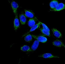
ARG43134 anti-FLCN antibody ICC/IF image
Immunofluorescence: U2OS cells were blocked with 10% goat serum and then stained with ARG43134 anti-FLCN antibody (green) at 2 µg/ml dilution, overnight at 4°C. DAPI (blue) for nuclear staining.
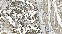
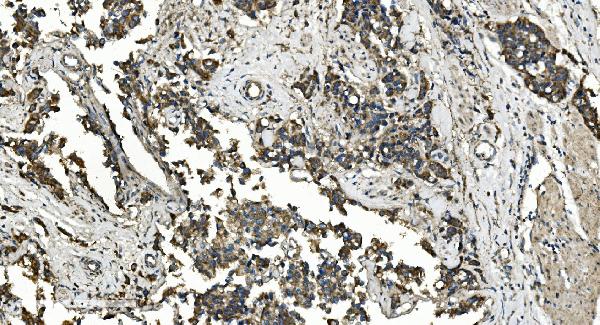
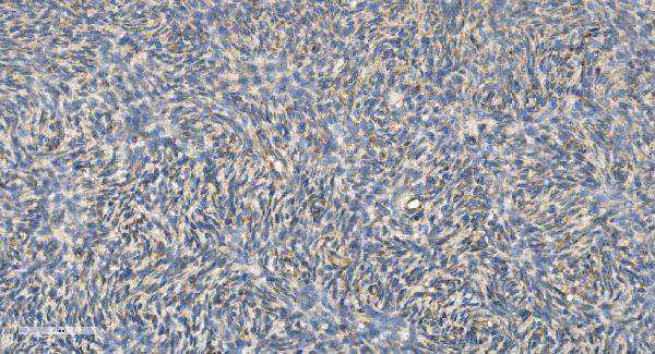
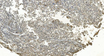
ARG43134 anti-FLCN antibody IHC-P image
Immunohistochemistry: Paraffin-embedded Human bladder cancer tissue. Antigen Retrieval: Heat mediation was performed in EDTA buffer (pH 8.0). The tissue section was blocked with 10% goat serum. The tissue section was then stained with ARG43134 anti-FLCN antibody at 1 µg/ml dilution, overnight at 4°C.

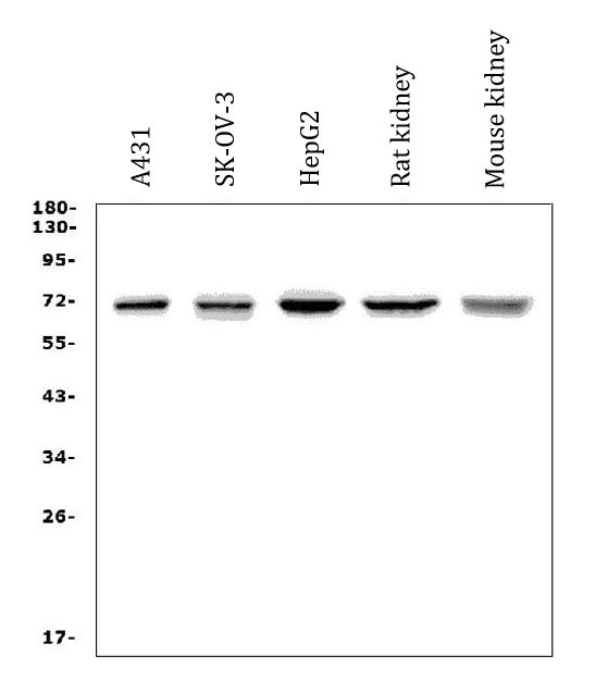
ARG43134 anti-FLCN antibody WB image
Western blot: 50 µg of sample under reducing conditions. A431, SK-OV-3, HepG2, Rat kidney and Mouse kidney lysates stained with ARG43134 anti-FLCN antibody at 0.5 µg/ml dilution, overnight at 4°C.
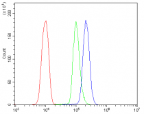
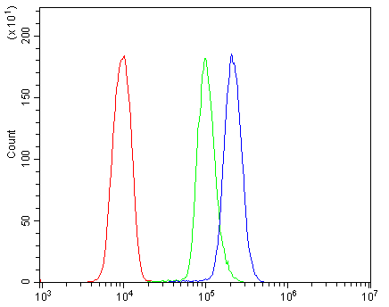
ARG43134 anti-FLCN antibody FACS image
Flow Cytometry: Caco-2 cells were blocked with 10% normal goat serum and then stained with ARG43134 anti-FLCN antibody (blue) at 1 µg/10^6 cells for 30 min at 20°C, followed by incubation with DyLight®488 labelled secondary antibody. Isotype control antibody (green) was rabbit IgG (1 µg/10^6 cells) used under the same conditions. Unlabelled sample (red) was also used as a control.
ARG43134 anti-FLCN antibody IHC-P image
Immunohistochemistry: Paraffin-embedded Human bladder cancer tissue. Antigen Retrieval: Heat mediation was performed in EDTA buffer (pH 8.0). The tissue section was blocked with 10% goat serum. The tissue section was then stained with ARG43134 anti-FLCN antibody at 1 µg/ml dilution, overnight at 4°C.
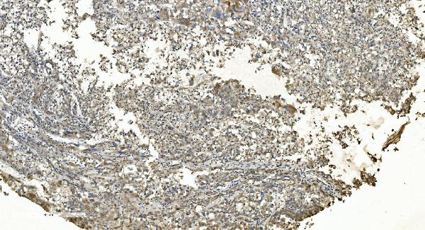
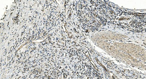
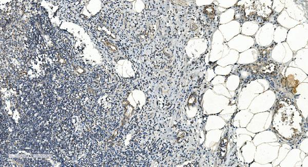
ARG43134 anti-FLCN antibody IHC-P image
Immunohistochemistry: Paraffin-embedded Human appendicitis tissue. Antigen Retrieval: Heat mediation was performed in EDTA buffer (pH 8.0). The tissue section was blocked with 10% goat serum. The tissue section was then stained with ARG43134 anti-FLCN antibody at 1 µg/ml dilution, overnight at 4°C.
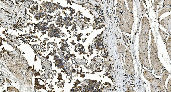
ARG43134 anti-FLCN antibody IHC-P image
Immunohistochemistry: Paraffin-embedded Human liver cancer tissue. Antigen Retrieval: Heat mediation was performed in EDTA buffer (pH 8.0). The tissue section was blocked with 10% goat serum. The tissue section was then stained with ARG43134 anti-FLCN antibody at 1 µg/ml dilution, overnight at 4°C.

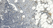
ARG43134 anti-FLCN antibody IHC-P image
Immunohistochemistry: Paraffin-embedded Human skin cancer tissue. Antigen Retrieval: Heat mediation was performed in EDTA buffer (pH 8.0). The tissue section was blocked with 10% goat serum. The tissue section was then stained with ARG43134 anti-FLCN antibody at 1 µg/ml dilution, overnight at 4°C.

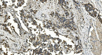
ARG43134 anti-FLCN antibody IHC-P image
Immunohistochemistry: Paraffin-embedded Human rectal cancer tissue. Antigen Retrieval: Heat mediation was performed in EDTA buffer (pH 8.0). The tissue section was blocked with 10% goat serum. The tissue section was then stained with ARG43134 anti-FLCN antibody at 1 µg/ml dilution, overnight at 4°C.
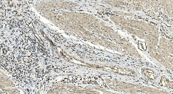
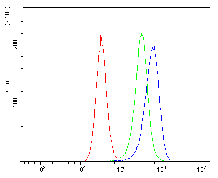
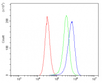
ARG43134 anti-FLCN antibody FACS image
Flow Cytometry: U937 cells were blocked with 10% normal goat serum and then stained with ARG43134 anti-FLCN antibody (blue) at 1 µg/10^6 cells for 30 min at 20°C, followed by incubation with DyLight®488 labelled secondary antibody. Isotype control antibody (green) was rabbit IgG (1 µg/10^6 cells) used under the same conditions. Unlabelled sample (red) was also used as a control.


 New Products


