


anti-GFP antibody


Key features and details
- 产品描述:
- 反应物种:
- 应用:
- 宿主:
- 克隆:
- 同位型:
- 靶点名称:
- 抗原物种:
- 抗原:
-
Brand:

 Product Details
| 产品描述 | Chicken Polyclonal Antibody to GFP |
|---|---|
| 反应物种 | Other |
| 应用 | ICC/IF, IHC-Fr, WB |
| 宿主 | Chicken |
| 克隆 | Polyclonal |
| 同位型 | IgY |
| 靶点名称 | GFP |
| 抗原物种 | Others |
| 抗原 | Prot-aceGFP recombinant protein. |
| 偶联标记 | Un-conjugated |
| 应用建议 |
| ||||||||
|---|---|---|---|---|---|---|---|---|---|
| 应用说明 | * The dilutions indicate recommended starting dilutions and the optimal dilutions or concentrations should be determined by the scientist. |
| 形式 | Liquid |
|---|---|
| 纯化 | Affinity purification. |
| 缓冲液 | PBS and 50% Glycerol. |
| 稳定剂 | 50% Glycerol |
| 浓度 | 1 mg/ml |
| 存放说明 | For continuous use, store undiluted antibody at 2-8°C for up to a week. For long-term storage, aliquot and store at -20°C. Storage in frost free freezers is not recommended. Avoid repeated freeze/thaw cycles. Suggest spin the vial prior to opening. The antibody solution should be gently mixed before use. |
| 注意事项 | For laboratory research only, not for drug, diagnostic or other use. |
| 研究领域 | Controls and Markers antibody; Tag Internal Control antibody; Fluorescent-Tags antibody |
|---|
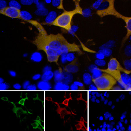
ARG10755 anti-GFP antibody ICC/IF image
Immunocytochemistry: Transfected Hek293 cells which overexpressed GFP-fusion protein fused to a nuclear localization sequence were stained with ARG10755 anti-GFP antibody (red). Most Hek293 cells are not transfected so only the nucleus of these cells can be visualized with a blue DNA stain. Cells which are transfected with GFP are bright green. Red antibody staining is only seen in cells which express GFP, as expected, and the superimposition of green and red results in an orange signal.

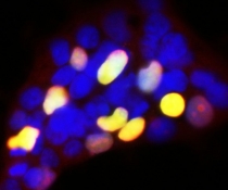
ARG10755 anti-GFP antibody ICC/IF image
Immunofluorescence: Transfected HEK293 cells with a GFP fusion protein construct (green), and stained with ARG10755 anti-GFP antibody (red) at 1:1000 dilution. DAPI (blue) for nuclear staining.
The GFP antibody reveals GFP protein expressed only in transfected cells, and as a result these cells appear golden yellow in color. Top, merged image, bottom individual channels.
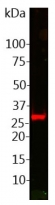
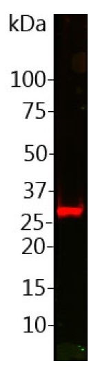
ARG10755 anti-GFP antibody WB image
Western blot: HEK293 cells transfected with pFin-EF1-GFP vector. This vector expresses full length GFP and crude homogenate from these cells was stained with ARG10755 anti-GFP antibody.
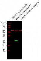
ARG10755 anti-GFP antibody WB image
Western blot: 1) HEK293 (non-transfected), 2) HEK293 transfected with a GFP construct and 3) HEK293 transfected with an mCherry construct. The blots were stained with ARG10755 anti-GFP antibody (green) at 1:1000 dilution. The blot was simultaneously stained with ARG10757 anti-Hsp 60 antibody (red) at 1:10000 dilution.

 New Products


