


anti-HLA C antibody


Key features and details
- 产品描述:
- 反应物种:
- 应用:
- 宿主:
- 克隆:
- 同位型:
- 靶点名称:
- 抗原物种:
- 抗原:
-
Brand:

 Product Details
| 产品描述 | Rabbit Polyclonal antibody recognizes HLA C |
|---|---|
| 反应物种 | Hu |
| 应用 | FACS, ICC, IHC-P, WB |
| 宿主 | Rabbit |
| 克隆 | Polyclonal |
| 同位型 | IgG |
| 靶点名称 | HLA C |
| 抗原物种 | Human |
| 抗原 | Synthetic peptide corresponding to aa. 38-62 of Human HLA C. (EYWDRETQKYKRQAQADRVNLRKLR) |
| 偶联标记 | Un-conjugated |
| 別名 | HLA-JY3; PSORS1; D6S204; MHC class I antigen Cw*7; HLC-C; HLA class I histocompatibility antigen, Cw-7 alpha chain |
| 应用建议 |
| ||||||||||
|---|---|---|---|---|---|---|---|---|---|---|---|
| 应用说明 | IHC-P: Antigen Retrieval: Heat mediation was performed in Citrate buffer (pH 6.0, epitope retrieval solution) for 20 min. * The dilutions indicate recommended starting dilutions and the optimal dilutions or concentrations should be determined by the scientist. | ||||||||||
| 阳性对照 | HeLa, A549 and U87 | ||||||||||
| 实际分子量 | ~ 41 kDa |
| 形式 | Liquid |
|---|---|
| 纯化 | Affinity purification with immunogen. |
| 缓冲液 | 0.2% Na2HPO4, 0.9% NaCl, 0.05% Sodium azide and 5% BSA. |
| 抗菌剂 | 0.05% Sodium azide |
| 稳定剂 | 5% BSA |
| 浓度 | 0.5 mg/ml |
| 存放说明 | For continuous use, store undiluted antibody at 2-8°C for up to a week. For long-term storage, aliquot and store at -20°C or below. Storage in frost free freezers is not recommended. Avoid repeated freeze/thaw cycles. Suggest spin the vial prior to opening. The antibody solution should be gently mixed before use. |
| 注意事项 | For laboratory research only, not for drug, diagnostic or other use. |
| 数据库连接 | Swiss-port # P10321 Human HLA class I histocompatibility antigen, Cw-7 alpha chain |
|---|---|
| 基因名称 | HLA-C |
| 全名 | major histocompatibility complex, class I, C |
| 生物功能 | Antigen-presenting major histocompatibility complex class I (MHCI) molecule with an important role in reproduction and antiviral immunity (PubMed:20972337, PubMed:24091323, PubMed:20439706, PubMed:11172028, PubMed:20104487, PubMed:28649982, PubMed:29312307). In complex with B2M/beta 2 microglobulin displays a restricted repertoire of self and viral peptides and acts as a dominant ligand for inhibitory and activating killer immunoglobulin receptors (KIRs) expressed on NK cells (PubMed:16141329). In an allogeneic setting, such as during pregnancy, mediates interaction of extravillous trophoblasts with KIR on uterine NK cells and regulate trophoblast invasion necessary for placentation and overall fetal growth (PubMed:20972337, PubMed:24091323). During viral infection, may present viral peptides with low affinity for KIRs, impeding KIR-mediated inhibition through peptide antagonism and favoring lysis of infected cells (PubMed:20439706). Presents a restricted repertoire of viral peptides on antigen-presenting cells for recognition by alpha-beta T cell receptor (TCR) on HLA-C-restricted CD8-positive T cells, guiding antigen-specific T cell immune response to eliminate infected cells, particularly in chronic viral infection settings such as HIV-1 or CMV infection (PubMed:11172028, PubMed:20104487, PubMed:28649982). Both the peptide and the MHC molecule are recognized by TCR, the peptide is responsible for the fine specificity of antigen recognition and MHC residues account for the MHC restriction of T cells (By similarity). Typically presents intracellular peptide antigens of 9 amino acids that arise from cytosolic proteolysis via proteasome. Can bind different peptides containing allele-specific binding motifs, which are mainly defined by anchor residues at position 2 and 9. Preferentially displays peptides having a restricted repertoire of hydrophobic or aromatic amino acids (Phe, Ile, Leu, Met, Val and Tyr) at the C-terminal anchor (PubMed:8265661, PubMed:25311805). ALLELE C*01:02: The peptide-bound form interacts with KIR2DL2 and KIR2DL3 inhibitory receptors on NK cells. The low affinity peptides compete with the high affinity peptides impeding KIR-mediated inhibition and favoring lysis of infected cells (PubMed:20439706). Presents to CD8-positive T cells a CMV epitope derived from UL83/pp65 (RCPEMISVL), an immediate-early antigen necessary for initiating viral replication (PubMed:12947002). ALLELE C*04:01: Presents a conserved HIV-1 epitope derived from env (SFNCGGEFF) to memory CD8-positive T cells, eliciting very strong IFNG responses (PubMed:20104487). Presents CMV epitope derived from UL83/pp65 (QYDPVAALF) to CD8-positive T cells, triggering T cell cytotoxic response (PubMed:12947002). ALLELE C*05:01: Presents HIV-1 epitope derived from rev (SAEPVPLQL) to CD8-positive T cells, triggering T cell cytotoxic response. ALLELE C*06:02: In trophoblasts, interacts with KIR2DS2 on uterine NK cells and triggers NK cell activation, including secretion of cytokines such as GMCSF that enhances trophoblast migration. ALLELE C*07:02: Plays an important role in the control of chronic CMV infection. Presents immunodominant CMV epitopes derived from IE1 (LSEFCRVL and CRVLCCYVL) and UL28 (FRCPRRFCF), both antigens synthesized during immediate-early period of viral replication. Elicits a strong anti-viral CD8-positive T cell immune response that increases markedly with age. ALLELE C*08:01: Presents viral epitopes derived from CMV UL83 (VVCAHELVC) and IAV M1 (GILGFVFTL), triggering CD8-positive T cell cytotoxic response. ALLELE C*12:02: Presents CMV epitope derived from UL83 (VAFTSHEHF) to CD8-positive T cells. ALLELE C*15:02: Presents CMV epitope derived from UL83 CC (VVCAHELVC) to CD8-positive T cells, triggering T cell cytotoxic response. [UniProt] |
| 细胞定位 | Membrane; Single-pass type I membrane protein. [UniProt] |
| 预测分子量 | 41 kDa |
| 翻译后修饰 | Polyubiquitinated in a post ER compartment by interaction with human herpesvirus 8 MIR1 protein. This targets the protein for rapid degradation via the ubiquitin system (By similarity). [UniProt] |

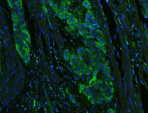
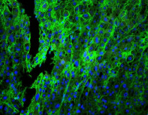
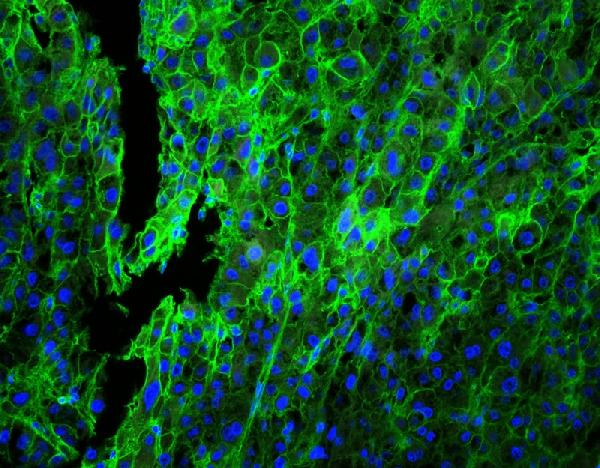
ARG42194 anti-HLA C antibody ICC image
Immunocytochemistry: HeLa cells stained with ARG42194 anti-HLA C antibody at 1 µg/ml dilution, overnight at 4°C.

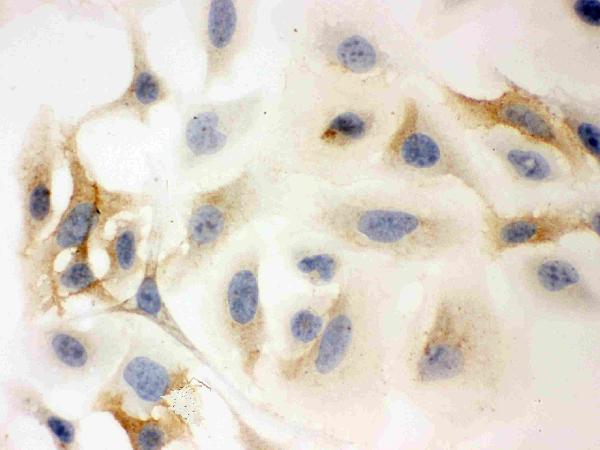
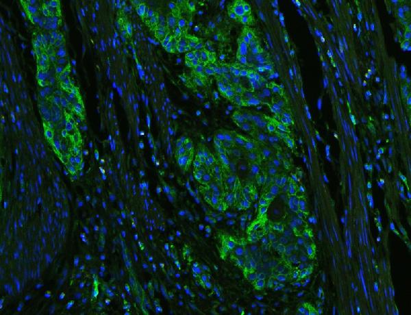
ARG42194 anti-HLA C antibody IHC-P image
Immunohistochemistry: Paraffin-embedded Human lung cancer tissue. Antigen Retrieval: Heat mediation was performed in Citrate buffer (pH 6.0, epitope retrieval solution) for 20 min. The tissue section was blocked with 10% goat serum. The tissue section was then stained with ARG42194 anti-HLA C antibody at 1 µg/ml dilution, overnight at 4°C.

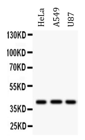
ARG42194 anti-HLA C antibody WB image
Western blot: 50 µg of samples under reducing conditions. HeLa, A549 and U87 whole cell lysates stained with ARG42194 anti-HLA C antibody at 0.5 µg/ml dilution, overnight at 4°C.
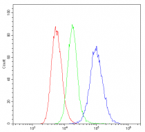
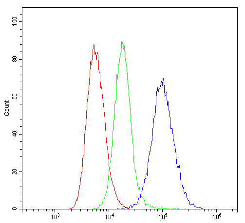
ARG42194 anti-HLA C antibody FACS image
Flow Cytometry: SiHa cells were blocked with 10% normal goat serum and then stained with ARG42194 anti-HLA C antibody (blue line) at 1 µg/10^6 cells for 30 min at 20°C, followed by incubation with DyLight®488 labelled secondary antibody. Isotype control antibody (green line) was Rabbit IgG (1 µg/10^6 cells) used under the same conditions. Unlabelled sample (red line) was also used as a control.
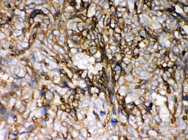
ARG42194 anti-HLA C antibody IHC-P image
Immunohistochemistry: Paraffin-embedded Human lung cancer tissue. Antigen Retrieval: Heat mediation was performed in Citrate buffer (pH 6.0, epitope retrieval solution) for 20 min. The tissue section was blocked with 10% goat serum. The tissue section was then stained with ARG42194 anti-HLA C antibody at 1 µg/ml dilution, overnight at 4°C.
ARG42194 anti-HLA C antibody IHC-P image
Immunohistochemistry: Paraffin-embedded Human oesophagus squama cancer tissue. Antigen Retrieval: Heat mediation was performed in Citrate buffer (pH 6.0, epitope retrieval solution) for 20 min. The tissue section was blocked with 10% goat serum. The tissue section was then stained with ARG42194 anti-HLA C antibody at 1 µg/ml dilution, overnight at 4°C.


 New Products


