


anti-HOOK2 antibody


Key features and details
- 产品描述:
- 反应物种:
- 应用:
- 宿主:
- 克隆:
- 同位型:
- 靶点名称:
- 抗原物种:
- 抗原:
-
Brand:

 Product Details
| 产品描述 | Rabbit Polyclonal antibody recognizes HOOK2 |
|---|---|
| 反应物种 | Hu, Ms, Rat |
| 应用 | FACS, ICC/IF, IHC-P, WB |
| 宿主 | Rabbit |
| 克隆 | Polyclonal |
| 同位型 | IgG |
| 靶点名称 | HOOK2 |
| 抗原物种 | Human |
| 抗原 | Recombinant protein corresponding to E500-R702 of Human HOOK2. |
| 偶联标记 | Un-conjugated |
| 別名 | HK2; hHK2; Protein Hook homolog 2; h-hook2 |
| 应用建议 |
| ||||||||||
|---|---|---|---|---|---|---|---|---|---|---|---|
| 应用说明 | IHC-P: Antigen Retrieval: Heat mediation was performed in Citrate buffer (pH 6.0) for 20 min. * The dilutions indicate recommended starting dilutions and the optimal dilutions or concentrations should be determined by the scientist. | ||||||||||
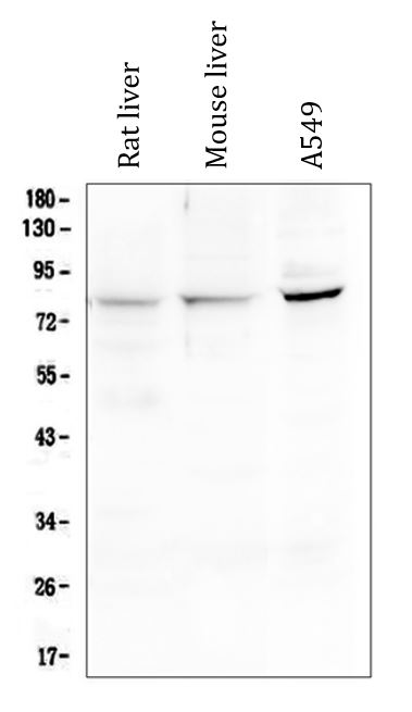
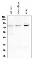
| 阳性对照 | Rat liver, Mouse liver and A549 | ||||||||||
| 实际分子量 | ~ 83 kDa |
| 形式 | Liquid |
|---|---|
| 纯化 | Affinity purification with immunogen. |
| 缓冲液 | 0.2% Na2HPO4, 0.9% NaCl, 0.05% Sodium azide and 4% Trehalose. |
| 抗菌剂 | 0.05% Sodium azide |
| 稳定剂 | 4% Trehalose |
| 浓度 | 0.5 mg/ml |
| 存放说明 | For continuous use, store undiluted antibody at 2-8°C for up to a week. For long-term storage, aliquot and store at -20°C or below. Storage in frost free freezers is not recommended. Avoid repeated freeze/thaw cycles. Suggest spin the vial prior to opening. The antibody solution should be gently mixed before use. |
| 注意事项 | For laboratory research only, not for drug, diagnostic or other use. |
| 数据库连接 | |
|---|---|
| 基因名称 | HOOK2 |
| 全名 | hook microtubule-tethering protein 2 |
| 背景介绍 | Hook proteins are cytosolic coiled-coil proteins that contain conserved N-terminal domains, which attach to microtubules, and more divergent C-terminal domains, which mediate binding to organelles. The Drosophila Hook protein is a component of the endocytic compartment.[supplied by OMIM, Apr 2004] |
| 生物功能 | Component of the FTS/Hook/FHIP complex (FHF complex). The FHF complex may function to promote vesicle trafficking and/or fusion via the homotypic vesicular protein sorting complex (the HOPS complex). Contributes to the establishment and maintenance of centrosome function. May function in the positioning or formation of aggresomes, which are pericentriolar accumulations of misfolded proteins, proteasomes and chaperones. [UniProt] |
| 细胞定位 | Cytoplasm, cytoskeleton, microtubule organizing center, centrosome. Note=Colocalizes with aggresomes, which are aggregates of misfolded proteins, at the centrosome. Also localizes to punctate cytoplasmic foci which do not appear to overlap with early or late endosomes, the endoplasmic reticulum, the Golgi complex, multivesicular bodies (MVBs), lysosome, or mitochondria. Often found in close association with microtubules. Localizes to the manchette in elongating spermatids. [UniProt] |
| 预测分子量 | 83 kDa |

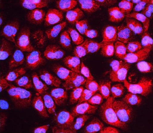
ARG43093 anti-HOOK2 antibody ICC/IF image
Immunofluorescence: A431 cells were blocked with 10% goat serum and then stained with ARG43093 anti-HOOK2 antibody (red) at 2 µg/ml dilution, overnight at 4°C. DAPI (blue) for nuclear staining.
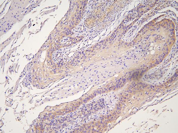
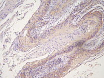
ARG43093 anti-HOOK2 antibody IHC-P image
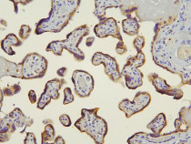
Immunohistochemistry: Paraffin-embedded Human placenta tissue. Antigen Retrieval: Heat mediation was performed in Citrate buffer (pH 6.0) for 20 min. The tissue section was blocked with 10% goat serum. The tissue section was then stained with ARG43093 anti-HOOK2 antibody at 1 µg/ml dilution, overnight at 4°C.
ARG43093 anti-HOOK2 antibody WB image
Western blot: 50 µg of sample under reducing conditions. Rat liver, Mouse liver and A549 whole cell lysates stained with ARG43093 anti-HOOK2 antibody at 0.5 µg/ml dilution, overnight at 4°C.
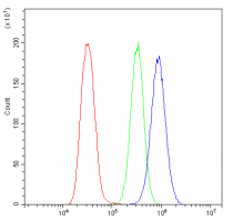
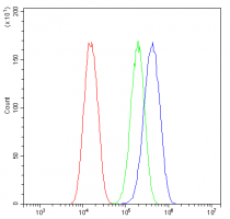
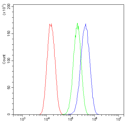
ARG43093 anti-HOOK2 antibody FACS image
Flow Cytometry: U2OS cells were blocked with 10% normal goat serum and then stained with ARG43093 anti-HOOK2 antibody (blue) at 1 µg/10^6 cells for 30 min at 20°C, followed by incubation with DyLight®488 labelled secondary antibody. Isotype control antibody (green) was rabbit IgG (1 µg/10^6 cells) used under the same conditions. Unlabelled sample (red) was also used as a control.
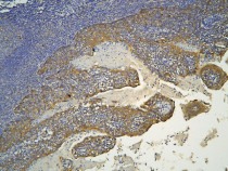
ARG43093 anti-HOOK2 antibody IHC-P image
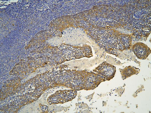
Immunohistochemistry: Paraffin-embedded Human oesophagus squama cancer tissue. Antigen Retrieval: Heat mediation was performed in Citrate buffer (pH 6.0) for 20 min. The tissue section was blocked with 10% goat serum. The tissue section was then stained with ARG43093 anti-HOOK2 antibody at 1 µg/ml dilution, overnight at 4°C.
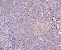
ARG43093 anti-HOOK2 antibody IHC-P image
Immunohistochemistry: Paraffin-embedded Human sarcoma tissue. Antigen Retrieval: Heat mediation was performed in Citrate buffer (pH 6.0) for 20 min. The tissue section was blocked with 10% goat serum. The tissue section was then stained with ARG43093 anti-HOOK2 antibody at 1 µg/ml dilution, overnight at 4°C.
ARG43093 anti-HOOK2 antibody IHC-P image
Immunohistochemistry: Paraffin-embedded Human tonsil tissue. Antigen Retrieval: Heat mediation was performed in Citrate buffer (pH 6.0) for 20 min. The tissue section was blocked with 10% goat serum. The tissue section was then stained with ARG43093 anti-HOOK2 antibody at 1 µg/ml dilution, overnight at 4°C.

ARG43093 anti-HOOK2 antibody IHC-P image
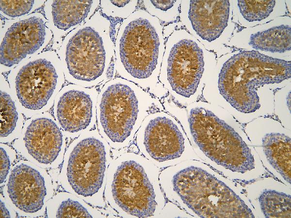
Immunohistochemistry: Paraffin-embedded Rat testis tissue. Antigen Retrieval: Heat mediation was performed in Citrate buffer (pH 6.0) for 20 min. The tissue section was blocked with 10% goat serum. The tissue section was then stained with ARG43093 anti-HOOK2 antibody at 1 µg/ml dilution, overnight at 4°C.
ARG43093 anti-HOOK2 antibody FACS image
Flow Cytometry: U87 cells were blocked with 10% normal goat serum and then stained with ARG43093 anti-HOOK2 antibody (blue) at 1 µg/10^6 cells for 30 min at 20°C, followed by incubation with DyLight®488 labelled secondary antibody. Isotype control antibody (green) was rabbit IgG (1 µg/10^6 cells) used under the same conditions. Unlabelled sample (red) was also used as a control.


 New Products


