


anti-IDH1 antibody [16H7]
![anti-IDH1 antibody [16H7]](/upload/image/products/ARG42111_WB_2_210_205.jpg)
![anti-IDH1 antibody [16H7]](/upload/image/products/ARG42111_IF_1.jpg)
![anti-IDH1 antibody [16H7]](/upload/image/products/ARG42111_WB_1.jpg)
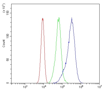
![anti-IDH1 antibody [16H7]](/upload/image/products/ARG42111_FACS_210707_1.jpg)
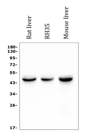
![anti-IDH1 antibody [16H7]](/upload/image/products/ARG42111_WB_2.jpg)
Key features and details
- 产品描述:
- 反应物种:
- 应用:
- 宿主:
- 克隆:
- 克隆号:
- 同位型:
- 靶点名称:
- 抗原物种:
-
Brand:

 Product Details
| 产品描述 | Mouse Monoclonal antibody [16H7] recognizes IDH1 |
|---|---|
| 反应物种 | Hu, Ms, Rat |
| 应用 | FACS, ICC/IF, WB |
| 宿主 | Mouse |
| 克隆 | Monoclonal |
| 克隆号 | 16H7 |
| 同位型 | IgG |
| 靶点名称 | IDH1 |
| 抗原物种 | Human |
| 抗原 | Synthetic peptide corresponding to aa. 381-413 of Human IDH1. (KGLPNVQRSDYLNTFEFMDKLGENLKIKLAQAK) |
| 偶联标记 | Un-conjugated |
| 別名 | IDPC; EC 1.1.1.42; Cytosolic NADP-isocitrate dehydrogenase; IDP; HEL-S-26; HEL-216; Isocitrate dehydrogenase [NADP] cytoplasmic; IDH; PICD; IDCD; NADP; Oxalosuccinate decarboxylase |
| 应用建议 |
| ||||||||
|---|---|---|---|---|---|---|---|---|---|
| 应用说明 | * The dilutions indicate recommended starting dilutions and the optimal dilutions or concentrations should be determined by the scientist. | ||||||||
| 实际分子量 | ~ 47 kDa |
| 形式 | Liquid |
|---|---|
| 纯化 | Affinity purification with immunogen. |
| 纯度 | > 95% (by SDS-PAGE) |
| 缓冲液 | 0.2% Na2HPO4, 0.9% NaCl, 0.05% Sodium azide and 4% Trehalose. |
| 抗菌剂 | 0.05% Sodium azide |
| 稳定剂 | 4% Trehalose |
| 浓度 | 0.5 mg/ml |
| 存放说明 | For continuous use, store undiluted antibody at 2-8°C for up to a week. For long-term storage, aliquot and store at -20°C or below. Storage in frost free freezers is not recommended. Avoid repeated freeze/thaw cycles. Suggest spin the vial prior to opening. The antibody solution should be gently mixed before use. |
| 注意事项 | For laboratory research only, not for drug, diagnostic or other use. |
| 数据库连接 | |
|---|---|
| 基因名称 | IDH1 |
| 全名 | isocitrate dehydrogenase 1 (NADP+), soluble |
| 背景介绍 | Isocitrate dehydrogenases catalyze the oxidative decarboxylation of isocitrate to 2-oxoglutarate. These enzymes belong to two distinct subclasses, one of which utilizes NAD(+) as the electron acceptor and the other NADP(+). Five isocitrate dehydrogenases have been reported: three NAD(+)-dependent isocitrate dehydrogenases, which localize to the mitochondrial matrix, and two NADP(+)-dependent isocitrate dehydrogenases, one of which is mitochondrial and the other predominantly cytosolic. Each NADP(+)-dependent isozyme is a homodimer. The protein encoded by this gene is the NADP(+)-dependent isocitrate dehydrogenase found in the cytoplasm and peroxisomes. It contains the PTS-1 peroxisomal targeting signal sequence. The presence of this enzyme in peroxisomes suggests roles in the regeneration of NADPH for intraperoxisomal reductions, such as the conversion of 2, 4-dienoyl-CoAs to 3-enoyl-CoAs, as well as in peroxisomal reactions that consume 2-oxoglutarate, namely the alpha-hydroxylation of phytanic acid. The cytoplasmic enzyme serves a significant role in cytoplasmic NADPH production. Alternatively spliced transcript variants encoding the same protein have been found for this gene. [provided by RefSeq, Sep 2013] |
| 细胞定位 | Cytoplasm. Peroxisome. [UniProt] |
| 产品亮点 | Related products: Isocitrate Dehydrogenase antibodies; Isocitrate Dehydrogenase ELISA Kits; Anti-Mouse IgG secondary antibodies; Related news: TCA intermediate fumarate promotes mitobiogenesis |
| 预测分子量 | 47 kDa |
| 翻译后修饰 | Acetylation at Lys-374 dramatically reduces catalytic activity. [UniProt] |
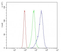
ARG42111 anti-IDH1 antibody [16H7] ICC/IF image
Immunofluorescence: U2OS cells stained with ARG42111 anti-IDH1 antibody [16H7] (green) at 2 µg/ml dilution, overnight at 4°C. DAPI (blue) for nuclear staining.

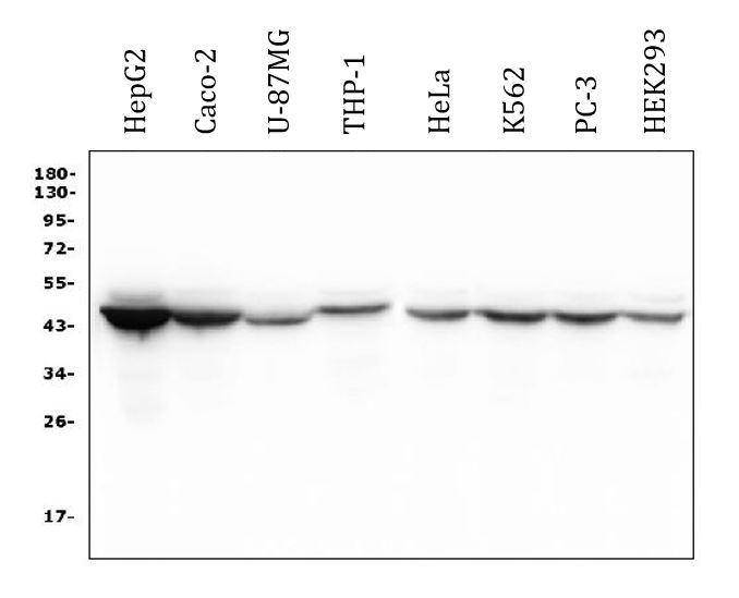
ARG42111 anti-IDH1 antibody [16H7] WB image
Western blot: 50 µg of samples under reducing conditions. HepG2, Caco-2, U-87MG, THP-1, HeLa, K562, PC-3 and HEK293 whole cell lysates stained with ARG42111 anti-IDH1 antibody [16H7] at 0.5 µg/ml dilution, overnight at 4°C.
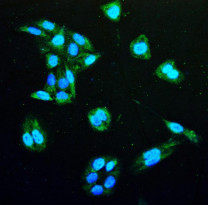
ARG42111 anti-IDH1 antibody [16H7] FACS image
Flow Cytometry: Caco-2 cells were blocked with 10% normal goat serum and then stained with ARG42111 anti-IDH1 antibody [16H7] (blue) at 1 µg/10^6 cells for 30 min at 20°C, followed by incubation with DyLight®488 labelled secondary antibody. Isotype control antibody (green) was mouse IgG (1 µg/10^6 cells) used under the same conditions. Unlabelled sample (red) was also used as a control.
ARG42111 anti-IDH1 antibody [16H7] WB image
Western blot: 50 µg of samples under reducing conditions. Rat liver, Rat RH35 and Mouse liver lysates stained with ARG42111 anti-IDH1 antibody [16H7] at 0.5 µg/ml dilution, overnight at 4°C.
 New Products


