


anti-MIF antibody


Key features and details
- 产品描述:
- 反应物种:
- 应用:
- 特异性:
- 宿主:
- 克隆:
- 同位型:
- 靶点名称:
- 抗原物种:
-
Brand:

 Product Details
| 产品描述 | Rabbit Polyclonal antibody recognizes MIF |
|---|---|
| 反应物种 | Hu, Ms, Rat |
| 应用 | IHC-P, WB |
| 特异性 | The antibody detects endogenous MIF protein. |
| 宿主 | Rabbit |
| 克隆 | Polyclonal |
| 同位型 | IgG |
| 靶点名称 | MIF |
| 抗原物种 | Human |
| 抗原 | Synthetic peptide around the Internal region of Human MIF. |
| 偶联标记 | Un-conjugated |
| 別名 | Phenylpyruvate tautomerase; L-dopachrome tautomerase; GLIF; L-dopachrome isomerase; MMIF; Macrophage migration inhibitory factor; MIF; GIF; EC 5.3.2.1; Glycosylation-inhibiting factor; EC 5.3.3.12 |
| 应用建议 |
| ||||||
|---|---|---|---|---|---|---|---|
| 应用说明 | IHC-P: Antigen Retrieval: Boil tissue section in Sodium citrate buffer (pH 6.0) for 20 min. * The dilutions indicate recommended starting dilutions and the optimal dilutions or concentrations should be determined by the scientist. |
| 形式 | Liquid |
|---|---|
| 纯化 | Affinity purification with immunogen. |
| 缓冲液 | PBS, 0.02% Sodium azide, 50% Glycerol and 0.5% BSA. |
| 抗菌剂 | 0.02% Sodium azide |
| 稳定剂 | 50% Glycerol and 0.5% BSA |
| 浓度 | 1 mg/ml |
| 存放说明 | For continuous use, store undiluted antibody at 2-8°C for up to a week. For long-term storage, aliquot and store at -20°C. Storage in frost free freezers is not recommended. Avoid repeated freeze/thaw cycles. Suggest spin the vial prior to opening. The antibody solution should be gently mixed before use. |
| 注意事项 | For laboratory research only, not for drug, diagnostic or other use. |
| 数据库连接 | |
|---|---|
| 基因名称 | MIF |
| 全名 | macrophage migration inhibitory factor (glycosylation-inhibiting factor) |
| 背景介绍 | This gene encodes a lymphokine involved in cell-mediated immunity, immunoregulation, and inflammation. It plays a role in the regulation of macrophage function in host defense through the suppression of anti-inflammatory effects of glucocorticoids. This lymphokine and the JAB1 protein form a complex in the cytosol near the peripheral plasma membrane, which may indicate an additional role in integrin signaling pathways. [provided by RefSeq, Jul 2008] |
| 生物功能 | Pro-inflammatory cytokine. Involved in the innate immune response to bacterial pathogens. The expression of MIF at sites of inflammation suggests a role as mediator in regulating the function of macrophages in host defense. Counteracts the anti-inflammatory activity of glucocorticoids. Has phenylpyruvate tautomerase and dopachrome tautomerase activity (in vitro), but the physiological substrate is not known. It is not clear whether the tautomerase activity has any physiological relevance, and whether it is important for cytokine activity. [UniProt] |
| 预测分子量 | 12 kDa |
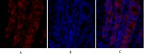
ARG66188 anti-MIF antibody IHC image
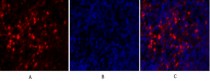
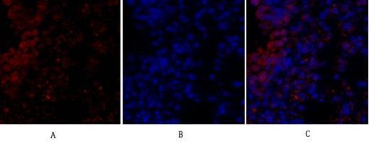
Immunohistochemistry: Rat lung tissue stained with ARG66188 anti-MIF antibody (red) at 1:200 dilution (4°C, overnight).
Picture A: Target. Picture B: DAPI. Picture C: merge of A+B.
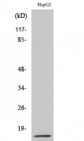
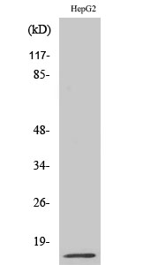
ARG66188 anti-MIF antibody WB image
Western blot: HepG2 cell lysate stained with ARG66188 anti-MIF antibody at 1:500 dilution.

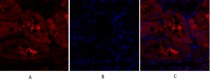
ARG66188 anti-MIF antibody IHC image
Immunohistochemistry: Rat lung tissue stained with ARG66188 anti-MIF antibody (red) at 1:200 dilution (4°C, overnight).
Picture A: Target. Picture B: DAPI. Picture C: merge of A+B.
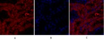
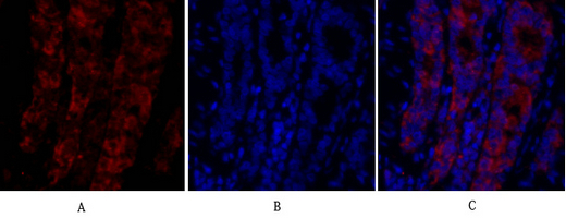
ARG66188 anti-MIF antibody IHC image
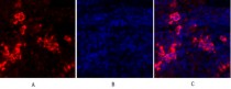
Immunohistochemistry: Rat kidney tissue stained with ARG66188 anti-MIF antibody (red) at 1:200 dilution (4°C, overnight).
Picture A: Target. Picture B: DAPI. Picture C: merge of A+B.
ARG66188 anti-MIF antibody IHC image
Immunohistochemistry: Rat kidney tissue stained with ARG66188 anti-MIF antibody (red) at 1:200 dilution (4°C, overnight).
Picture A: Target. Picture B: DAPI. Picture C: merge of A+B.
ARG66188 anti-MIF antibody IHC image
Immunohistochemistry: Rat spleen tissue stained with ARG66188 anti-MIF antibody (red) at 1:200 dilution (4°C, overnight).
Picture A: Target. Picture B: DAPI. Picture C: merge of A+B.
ARG66188 anti-MIF antibody IHC image
Immunohistochemistry: Rat spleen tissue stained with ARG66188 anti-MIF antibody (red) at 1:200 dilution (4°C, overnight).
Picture A: Target. Picture B: DAPI. Picture C: merge of A+B.
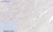
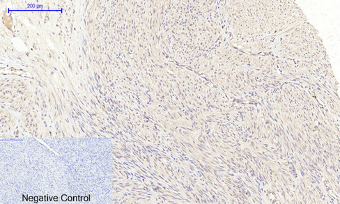
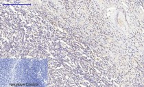
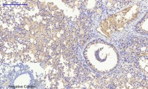
ARG66188 anti-MIF antibody IHC-P image
Immunohistochemistry: Paraffin-embedded Human uterus tissue stained with ARG66188 anti-MIF antibody at 1:200 dilution (4°C, overnight). Antigen Retrieval: Boil tissue section in Sodium citrate buffer (pH 6.0) for 20 min.
Negative control was used by secondary antibody only.
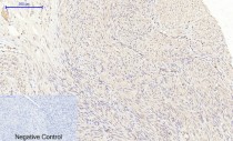
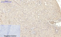
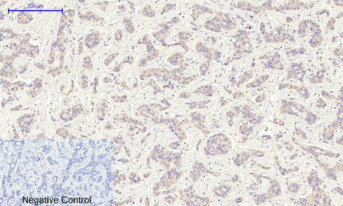
ARG66188 anti-MIF antibody IHC-P image
Immunohistochemistry: Paraffin-embedded Human uterus cancer tissue stained with ARG66188 anti-MIF antibody at 1:200 dilution (4°C, overnight). Antigen Retrieval: Boil tissue section in Sodium citrate buffer (pH 6.0) for 20 min.
Negative control was used by secondary antibody only.
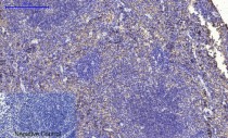
ARG66188 anti-MIF antibody IHC-P image
Immunohistochemistry: Paraffin-embedded Human Tonsil tissue stained with ARG66188 anti-MIF antibody at 1:200 dilution (4°C, overnight). Antigen Retrieval: Boil tissue section in Sodium citrate buffer (pH 6.0) for 20 min.
Negative control was used by secondary antibody only.
ARG66188 anti-MIF antibody IHC-P image
Immunohistochemistry: Paraffin-embedded Human liver tissue stained with ARG66188 anti-MIF antibody at 1:200 dilution (4°C, overnight). Antigen Retrieval: Boil tissue section in Sodium citrate buffer (pH 6.0) for 20 min.
Negative control was used by secondary antibody only.
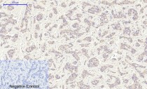
ARG66188 anti-MIF antibody IHC-P image
Immunohistochemistry: Paraffin-embedded Human liver cancer tissue stained with ARG66188 anti-MIF antibody at 1:200 dilution (4°C, overnight). Antigen Retrieval: Boil tissue section in Sodium citrate buffer (pH 6.0) for 20 min.
Negative control was used by secondary antibody only.

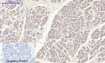
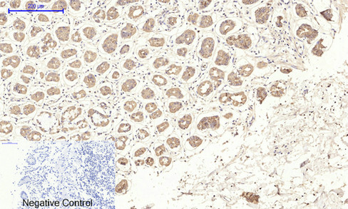
ARG66188 anti-MIF antibody IHC-P image
Immunohistochemistry: Paraffin-embedded Human stomach tissue stained with ARG66188 anti-MIF antibody at 1:200 dilution (4°C, overnight). Antigen Retrieval: Boil tissue section in Sodium citrate buffer (pH 6.0) for 20 min.
Negative control was used by secondary antibody only.
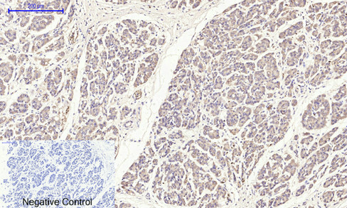
ARG66188 anti-MIF antibody IHC-P image
Immunohistochemistry: Paraffin-embedded Human stomach cancer tissue stained with ARG66188 anti-MIF antibody at 1:200 dilution (4°C, overnight). Antigen Retrieval: Boil tissue section in Sodium citrate buffer (pH 6.0) for 20 min.
Negative control was used by secondary antibody only.
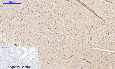
ARG66188 anti-MIF antibody IHC-P image
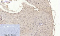
Immunohistochemistry: Paraffin-embedded Rat heart tissue stained with ARG66188 anti-MIF antibody at 1:200 dilution (4°C, overnight). Antigen Retrieval: Boil tissue section in Sodium citrate buffer (pH 6.0) for 20 min.
Negative control was used by secondary antibody only.

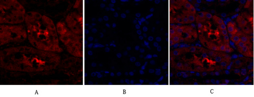
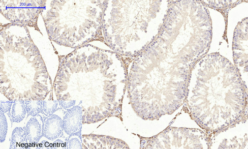
ARG66188 anti-MIF antibody IHC-P image
Immunohistochemistry: Paraffin-embedded Rat testis tissue stained with ARG66188 anti-MIF antibody at 1:200 dilution (4°C, overnight). Antigen Retrieval: Boil tissue section in Sodium citrate buffer (pH 6.0) for 20 min.
Negative control was used by secondary antibody only.
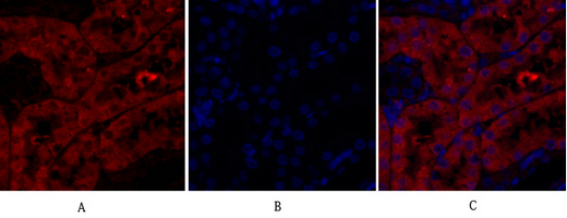
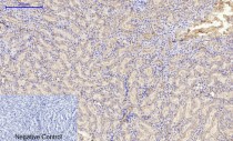
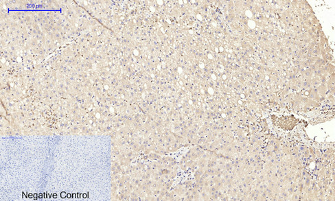
ARG66188 anti-MIF antibody IHC-P image
Immunohistochemistry: Paraffin-embedded Rat liver tissue stained with ARG66188 anti-MIF antibody at 1:200 dilution (4°C, overnight). Antigen Retrieval: Boil tissue section in Sodium citrate buffer (pH 6.0) for 20 min.
Negative control was used by secondary antibody only.

ARG66188 anti-MIF antibody IHC-P image
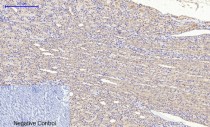
Immunohistochemistry: Paraffin-embedded Rat lung tissue stained with ARG66188 anti-MIF antibody at 1:200 dilution (4°C, overnight). Antigen Retrieval: Boil tissue section in Sodium citrate buffer (pH 6.0) for 20 min.
Negative control was used by secondary antibody only.
ARG66188 anti-MIF antibody IHC-P image
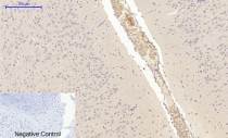
Immunohistochemistry: Paraffin-embedded Rat kidney tissue stained with ARG66188 anti-MIF antibody at 1:200 dilution (4°C, overnight). Antigen Retrieval: Boil tissue section in Sodium citrate buffer (pH 6.0) for 20 min.
Negative control was used by secondary antibody only.
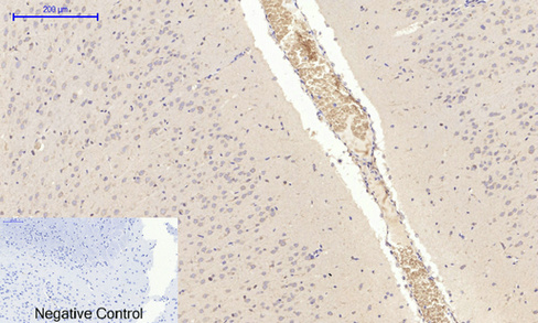
ARG66188 anti-MIF antibody IHC-P image
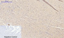
Immunohistochemistry: Paraffin-embedded Rat brain tissue stained with ARG66188 anti-MIF antibody at 1:200 dilution (4°C, overnight). Antigen Retrieval: Boil tissue section in Sodium citrate buffer (pH 6.0) for 20 min.
Negative control was used by secondary antibody only.
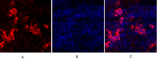
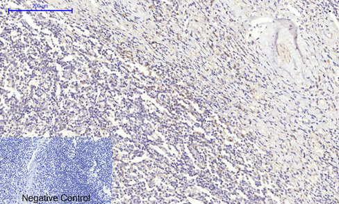
ARG66188 anti-MIF antibody IHC-P image
Immunohistochemistry: Paraffin-embedded Rat spleen tissue stained with ARG66188 anti-MIF antibody at 1:200 dilution (4°C, overnight). Antigen Retrieval: Boil tissue section in Sodium citrate buffer (pH 6.0) for 20 min.
Negative control was used by secondary antibody only.
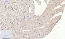
ARG66188 anti-MIF antibody IHC-P image
Immunohistochemistry: Paraffin-embedded Mouse heart tissue stained with ARG66188 anti-MIF antibody at 1:200 dilution (4°C, overnight). Antigen Retrieval: Boil tissue section in Sodium citrate buffer (pH 6.0) for 20 min.
Negative control was used by secondary antibody only.
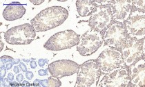
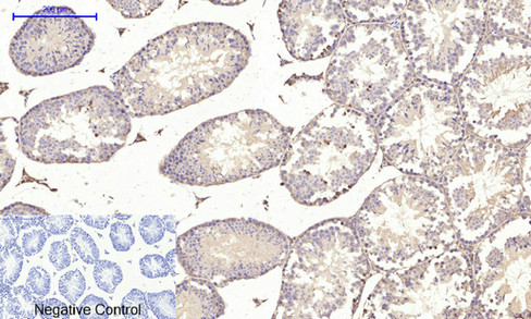
ARG66188 anti-MIF antibody IHC-P image
Immunohistochemistry: Paraffin-embedded Mouse testis tissue stained with ARG66188 anti-MIF antibody at 1:200 dilution (4°C, overnight). Antigen Retrieval: Boil tissue section in Sodium citrate buffer (pH 6.0) for 20 min.
Negative control was used by secondary antibody only.
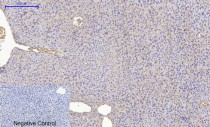
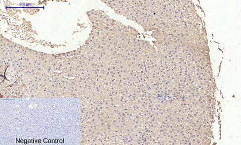
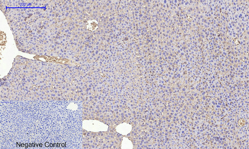
ARG66188 anti-MIF antibody IHC-P image
Immunohistochemistry: Paraffin-embedded Mouse liver tissue stained with ARG66188 anti-MIF antibody at 1:200 dilution (4°C, overnight). Antigen Retrieval: Boil tissue section in Sodium citrate buffer (pH 6.0) for 20 min.
Negative control was used by secondary antibody only.
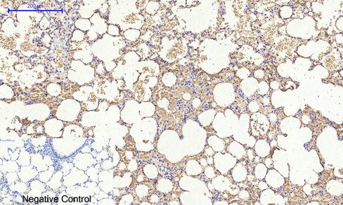
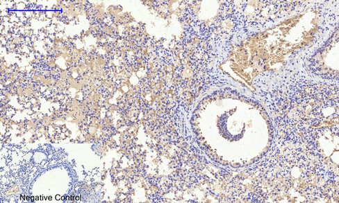
ARG66188 anti-MIF antibody IHC-P image
Immunohistochemistry: Paraffin-embedded Mouse lung tissue stained with ARG66188 anti-MIF antibody at 1:200 dilution (4°C, overnight). Antigen Retrieval: Boil tissue section in Sodium citrate buffer (pH 6.0) for 20 min.
Negative control was used by secondary antibody only.
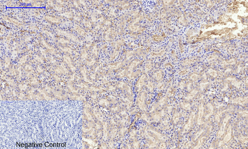
ARG66188 anti-MIF antibody IHC-P image
Immunohistochemistry: Paraffin-embedded Mouse kidney tissue stained with ARG66188 anti-MIF antibody at 1:200 dilution (4°C, overnight). Antigen Retrieval: Boil tissue section in Sodium citrate buffer (pH 6.0) for 20 min.
Negative control was used by secondary antibody only.
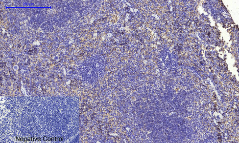
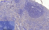
ARG66188 anti-MIF antibody IHC-P image
Immunohistochemistry: Paraffin-embedded Mouse spleen tissue stained with ARG66188 anti-MIF antibody at 1:200 dilution (4°C, overnight). Antigen Retrieval: Boil tissue section in Sodium citrate buffer (pH 6.0) for 20 min.
Negative control was used by secondary antibody only.


 New Products


