


anti-Neurofilament NF-H antibody

Key features and details
- 产品描述:
- 反应物种:
- 预测物种:
- 应用:
- 特异性:
- 宿主:
- 克隆:
- 同位型:
- 靶点名称:
-
Brand:

 Product Details
| 产品描述 | Chicken Polyclonal antibody recognizes Neurofilament NF-H |
|---|---|
| 反应物种 | Ms, Rat, Cow |
| 预测物种 | Hu, Chk |
| 应用 | ICC/IF, IHC-Fr, WB |
| 特异性 | The antibody recognizes phosphorylated NF-H strongly in samples. |
| 宿主 | Chicken |
| 克隆 | Polyclonal |
| 同位型 | IgY |
| 靶点名称 | Neurofilament NF-H |
| 抗原物种 | Bovine |
| 抗原 | Purified Bovine NF-H. |
| 偶联标记 | Un-conjugated |
| 別名 | Neurofilament heavy polypeptide; 200 kDa neurofilament protein; NF-H; Neurofilament triplet H protein; NFH |
| 应用建议 |
| ||||||||
|---|---|---|---|---|---|---|---|---|---|
| 应用说明 | Specific for the ~200k Neurofilament H protein. * The dilutions indicate recommended starting dilutions and the optimal dilutions or concentrations should be determined by the scientist. |
| 形式 | Liquid |
|---|---|
| 纯化 | Total IgY fraction |
| 缓冲液 | Total IgY fraction in PBS and 10 mM Sodium azide |
| 抗菌剂 | 10 mM Sodium azide |
| 存放说明 | For continuous use, store undiluted antibody at 2-8°C for up to a week. For long-term storage, aliquot and store at -20°C or below. Storage in frost free freezers is not recommended. Avoid repeated freeze/thaw cycles. Suggest spin the vial prior to opening. The antibody solution should be gently mixed before use. |
| 注意事项 | For laboratory research only, not for drug, diagnostic or other use. |
| 数据库连接 | |
|---|---|
| 背景介绍 | Neurofilaments are the 10nm or intermediate filament proteins found specifically in neurons, and are composed predominantly of three major proteins called NF-L, NF-M and NF-H . NF-H is the neurofilament high or heavy molecular weight polypeptide and runs on SDS-PAGE gels at 200-220 kDa, with some variability across species boundaries. Antibodies to NF-H are useful for identifying neuronal cells and their processes in tissue sections and in tissue culture. NF-H antibodies can also be useful to visualize neurofilament accumulations seen in many neurological diseases, such as Amyotrophic Lateral Sclerosis (Lou Gehrig's disease) (2) and Alzheimer's disease . |
| 产品亮点 | Related Antibody Duos and Panels: ARG30145 Intermediate Neurofilament Antibody Panel (NF-L, NF-M,NF-H) Related products: Neurofilament antibodies; Neurofilament ELISA Kits; Neurofilament Duos / Panels; Anti-Chicken IgY secondary antibodies; Related news: Neuronal Development Marker |
| 研究领域 | Neuroscience antibody; Signaling Transduction antibody; Neurofilament antibody; Intermediate Neurofilament antibody |
| 预测分子量 | 112 kDa |
| 翻译后修饰 | There are a number of repeats of the tripeptide K-S-P, NFH is phosphorylated on a number of the serines in this motif. It is thought that phosphorylation of NFH results in the formation of interfilament cross bridges that are important in the maintenance of axonal caliber. Phosphorylation seems to play a major role in the functioning of the larger neurofilament polypeptides (NF-M and NF-H), the levels of phosphorylation being altered developmentally and coincidentally with a change in the neurofilament function. Phosphorylated in the head and rod regions by the PKC kinase PKN1, leading to the inhibition of polymerization. |
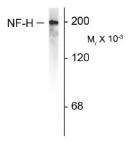
ARG52347 anti-Neurofilament NF-H antibody WB image
Western blot: Rat cortex lysate showing specific immunolableing of the ~200k NF-H protein stained with ARG52347 anti-Neurofilament NF-H antibody.

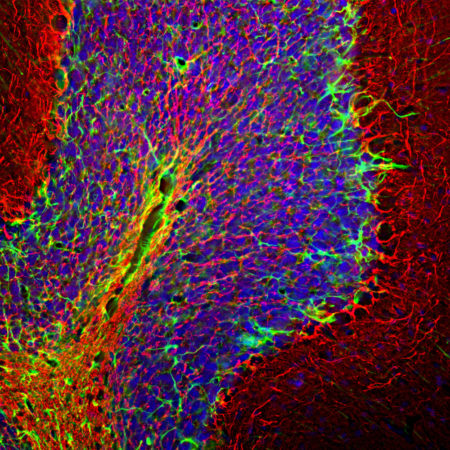
ARG52347 anti-Neurofilament NF-H antibody IHC-Fr image
Immunohistochemistry: Frozen section of Rat cerebellum tissue stained with ARG52347 anti-Neurofilament NF-H antibody (red) at 1:5000 dilution, and costained with anti-GFAP antibody (green) at 1:5000 dilution. DAPI (blue) for nuclear staining. Following transcardial perfusion with 4% paraformaldehyde, brain was post fixed for 24 hours, cut to 45 µM, and free floating sections were stained with above antibodies.
The NF-H antibody labels network of axons of different neurons, while the GFAP antibody stains astrocytes and other glial cells.
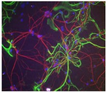
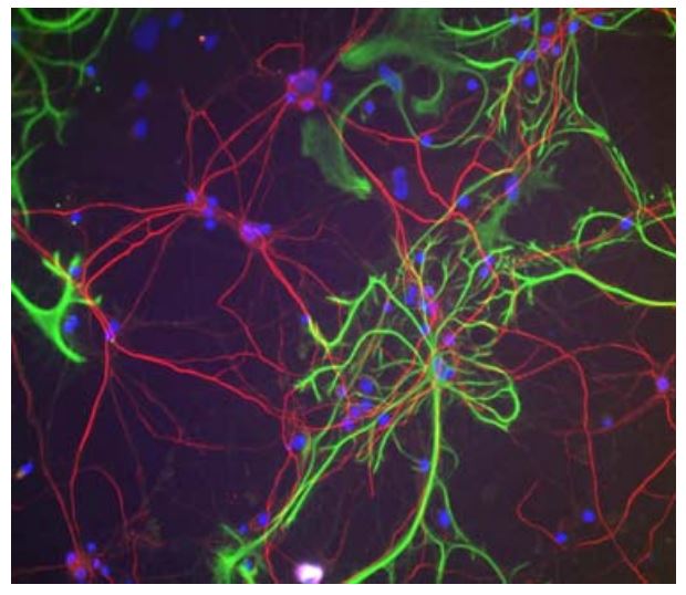
ARG52347 anti-Neurofilament NF-H antibody ICC/IF image
Immunofluorescence: Rat cortical neurons and glia stained with ARG52347 anti-Neurofilament NF-H antibody (red).
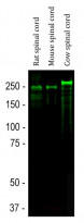
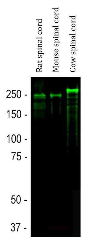
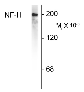
ARG52347 anti-Neurofilament NF-H antibody WB image
Western blot: Rat spinal cord, Mouse spinal cord and Cow spinal cord lysates stained with ARG52347 anti-Neurofilament NF-H antibody (green) at 1:20000 dilution.
Strong band at about 200-220 kDa corresponds to the phosphorylated from of NF-H. Smaller proteolytic fragments of NF-H are also detected in spinal cord preparations with this antibody.


 New Products


