


anti-NOVA2 antibody


Key features and details
- 产品描述:
- 反应物种:
- 应用:
- 宿主:
- 克隆:
- 同位型:
- 靶点名称:
- 抗原物种:
- 抗原:
-
Brand:

 Product Details
| 产品描述 | Rabbit Polyclonal antibody recognizes NOVA2 |
|---|---|
| 反应物种 | Hu, Ms, Rat |
| 应用 | FACS, IHC-P, WB |
| 宿主 | Rabbit |
| 克隆 | Polyclonal |
| 同位型 | IgG |
| 靶点名称 | NOVA2 |
| 抗原物种 | Human |
| 抗原 | Recombinant protein corresponding to M1-Q205 of Human NOVA2. |
| 偶联标记 | Un-conjugated |
| 別名 | NOVA3; RNA-binding protein Nova-2; Astrocytic NOVA1-like RNA-binding protein; ANOVA; Neuro-oncological ventral antigen 2 |
| 应用建议 |
| ||||||||
|---|---|---|---|---|---|---|---|---|---|
| 应用说明 | IHC-P: Antigen Retrieval: Heat mediation was performed in EDTA buffer (pH 8.0). * The dilutions indicate recommended starting dilutions and the optimal dilutions or concentrations should be determined by the scientist. | ||||||||
| 实际分子量 | 49, 72 kDa |
| 形式 | Liquid |
|---|---|
| 纯化 | Affinity purification with immunogen. |
| 缓冲液 | 0.2% Na2HPO4, 0.9% NaCl, 0.05% Sodium azide and 4% Trehalose. |
| 抗菌剂 | 0.05% Sodium azide |
| 稳定剂 | 4% Trehalose |
| 浓度 | 0.5 mg/ml |
| 存放说明 | For continuous use, store undiluted antibody at 2-8°C for up to a week. For long-term storage, aliquot and store at -20°C or below. Storage in frost free freezers is not recommended. Avoid repeated freeze/thaw cycles. Suggest spin the vial prior to opening. The antibody solution should be gently mixed before use. |
| 注意事项 | For laboratory research only, not for drug, diagnostic or other use. |
| 数据库连接 | |
|---|---|
| 基因名称 | NOVA2 |
| 全名 | neuro-oncological ventral antigen 2 |
| 生物功能 | May regulate RNA splicing or metabolism in a specific subset of developing neurons (By similarity). Binds single strand RNA. [UniProt] |
| 细胞定位 | Nucleus. [UniProt] |
| 预测分子量 | 49 kDa |
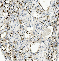
ARG42975 anti-NOVA2 antibody IHC-P image
Immunohistochemistry: Paraffin-embedded Human appendicitis tissue. Antigen Retrieval: Heat mediation was performed in EDTA buffer (pH 8.0). The tissue section was blocked with 10% goat serum. The tissue section was then stained with ARG42975 anti-NOVA2 antibody at 1 µg/ml dilution, overnight at 4°C.
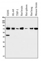
ARG42975 anti-NOVA2 antibody WB image
Western blot: 50 µg of sample under reducing conditions. U2OS, HL-60, THP-1, Rat brain, Rat spleen, Rat lung and Mouse brain lysates stained with ARG42975 anti-NOVA2 antibody at 0.5 µg/ml dilution, overnight at 4°C.
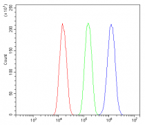
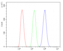
ARG42975 anti-NOVA2 antibody FACS image
Flow Cytometry: A549 cells were blocked with 10% normal goat serum and then stained with ARG42975 anti-NOVA2 antibody (blue) at 1 µg/10^6 cells for 30 min at 20°C, followed by incubation with DyLight®488 labelled secondary antibody. Isotype control antibody (green) was Rabbit IgG (1 µg/10^6 cells) used under the same conditions. Unlabelled sample (red) was also used as a control.

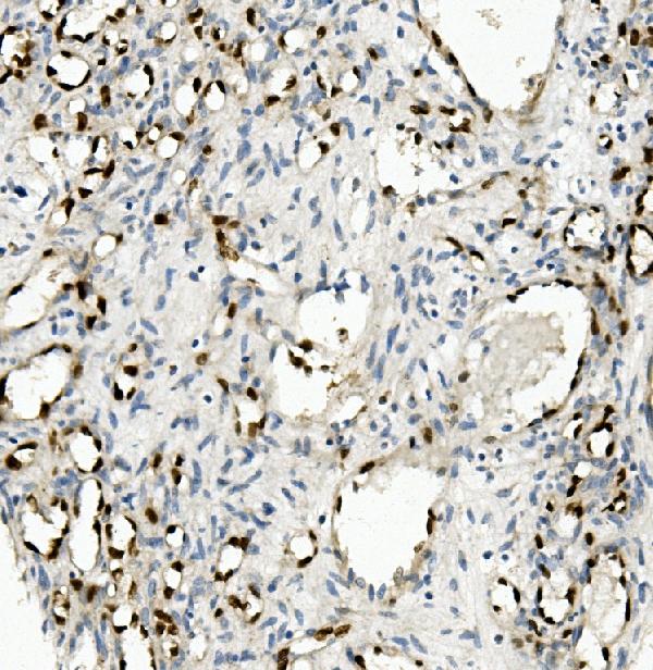
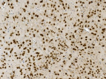
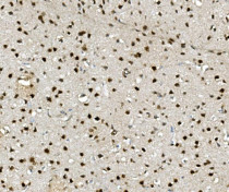
ARG42975 anti-NOVA2 antibody IHC-P image
Immunohistochemistry: Paraffin-embedded Mouse brain tissue. Antigen Retrieval: Heat mediation was performed in EDTA buffer (pH 8.0). The tissue section was blocked with 10% goat serum. The tissue section was then stained with ARG42975 anti-NOVA2 antibody at 1 µg/ml dilution, overnight at 4°C.
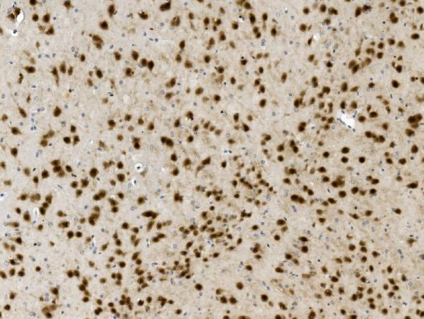
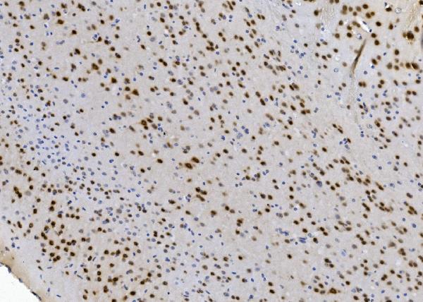
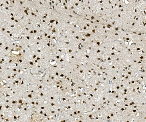
ARG42975 anti-NOVA2 antibody IHC-P image
Immunohistochemistry: Paraffin-embedded Rat liver tissue. Antigen Retrieval: Heat mediation was performed in EDTA buffer (pH 8.0). The tissue section was blocked with 10% goat serum. The tissue section was then stained with ARG42975 anti-NOVA2 antibody at 1 µg/ml dilution, overnight at 4°C.
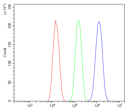
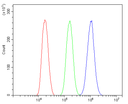
ARG42975 anti-NOVA2 antibody FACS image
Flow Cytometry: Hepa 1-6 cells were blocked with 10% normal goat serum and then stained with ARG42975 anti-NOVA2 antibody (blue) at 1 µg/10^6 cells for 30 min at 20°C, followed by incubation with DyLight®488 labelled secondary antibody. Isotype control antibody (green) was Rabbit IgG (1 µg/10^6 cells) used under the same conditions. Unlabelled sample (red) was also used as a control.


 New Products


