


anti-NQO1 antibody


Key features and details
- 产品描述:
- 反应物种:
- 预测物种:
- 应用:
- 特异性:
- 宿主:
- 克隆:
- 同位型:
- 靶点名称:
-
Brand:

 Product Details
| 产品描述 | Goat Polyclonal antibody recognizes NQO1 |
|---|---|
| 反应物种 | Hu, Rat, Pig |
| 预测物种 | Dog |
| 应用 | ICC/IF, IHC-P, WB |
| 特异性 | This products is expected to recognize all three reported isoforms (NP_000894.1,NP_001020604.1 and NP_001020605.1). |
| 宿主 | Goat |
| 克隆 | Polyclonal |
| 同位型 | IgG |
| 靶点名称 | NQO1 |
| 抗原物种 | Human |
| 抗原 | C-SIPTDNQIKARK |
| 偶联标记 | Un-conjugated |
| 別名 | NMOR1; EC 1.6.5.2; NAD; DHQU; P; Quinone reductase 1; DT-diaphorase; DTD; QR1; Phylloquinone reductase; NMORI; DIA4; Menadione reductase; Azoreductase |
| 应用建议 |
| ||||||||
|---|---|---|---|---|---|---|---|---|---|
| 应用说明 | WB: Recommend incubate at RT for 1h. IHC-P: Antigen Retrieval: Steam tissue section in Citrate buffer (pH 6.0). * The dilutions indicate recommended starting dilutions and the optimal dilutions or concentrations should be determined by the scientist. |
| 形式 | Liquid |
|---|---|
| 纯化 | Purified from goat serum by ammonium sulphate precipitation followed by antigen affinity chromatography using the immunizing peptide. |
| 缓冲液 | Tris saline (pH 7.3), 0.02% Sodium azide and 0.5% BSA |
| 抗菌剂 | 0.02% Sodium azide |
| 稳定剂 | 0.5% BSA |
| 浓度 | 0.5 mg/ml |
| 存放说明 | For continuous use, store undiluted antibody at 2-8°C for up to a week. For long-term storage, aliquot and store at -20°C or below. Storage in frost free freezers is not recommended. Avoid repeated freeze/thaw cycles. Suggest spin the vial prior to opening. The antibody solution should be gently mixed before use. |
| 注意事项 | For laboratory research only, not for drug, diagnostic or other use. |
| 数据库连接 | |
|---|---|
| 背景介绍 | This gene is a member of the NAD(P)H dehydrogenase (quinone) family and encodes a cytoplasmic 2-electron reductase. This FAD-binding protein forms homodimers and reduces quinones to hydroquinones. This protein's enzymatic activity prevents the one electron reduction of quinones that results in the production of radical species. Mutations in this gene have been associated with tardive dyskinesia (TD), an increased risk of hematotoxicity after exposure to benzene, and susceptibility to various forms of cancer. Altered expression of this protein has been seen in many tumors and is also associated with Alzheimer's disease (AD). Alternate transcriptional splice variants, encoding different isoforms, have been characterized. [provided by RefSeq, Jul 2008] |
| 产品亮点 | Related products: NQO1 antibodies; NQO1 Duos / Panels; Anti-Goat IgG secondary antibodies; Related news: Keap1-Nrf2-ARE antibody panel is launched |
| 研究领域 | Metabolism antibody; Signaling Transduction antibody |
| 预测分子量 | 31 kDa |
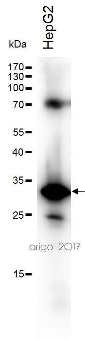
ARG63196 anti-NQO1 antibody WB image
Western blot: 30 µg of HepG2 cell lysate stained with ARG63196 anti-NQO1 antibody at 1:500 dilution.
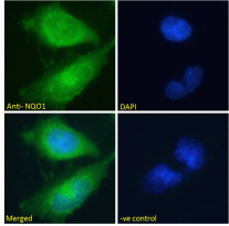
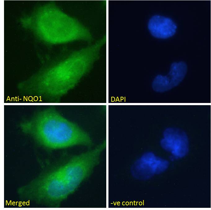
ARG63196 anti-NQO1 antibody ICC/IF image
Immunofluorescence: Paraformaldehyde fixed U251 cells permeabilized with 0.15% Triton. Cells were stained with ARG63196 anti-NQO1 antibody (green) at 10 µg/ml dilution for 1 hour. DAPI (blue) for nuclear staining. Negative control: Unimmunized goat IgG (green) at 10 µg/ml dilution.
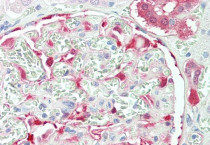
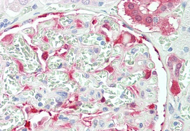
ARG63196 anti-NQO1 antibody IHC-P image
Immunohistochemistry: Paraffin-embedded Human kidney tissue. Antigen Retrieval: Steam tissue section in Citrate buffer (pH 6.0). The tissue section was stained with ARG63196 anti-NQO1 antibody at 5 µg/ml dilution followed by AP-staining.
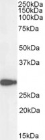
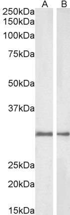
ARG63196 anti-NQO1 antibody WB image
Western blot: 35 µg of U251 cell lysate (in RIPA buffer) stained with ARG63196 anti-NQO1 antibody at 0.1 µg/ml dilution and incubated at RT for 1 hour.
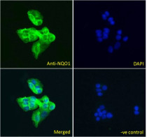
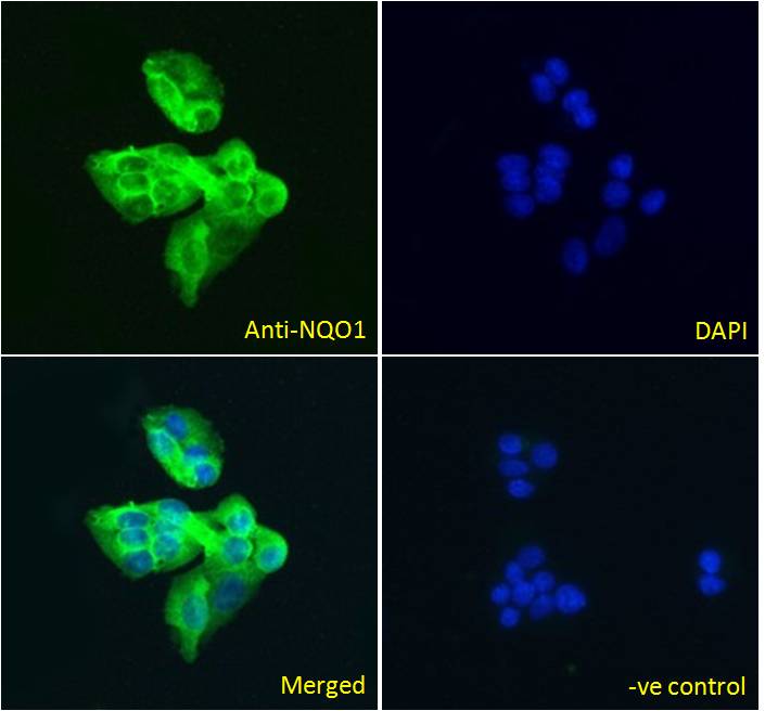
ARG63196 anti-NQO1 antibody ICC/IF image
Immunofluorescence: Paraformaldehyde fixed HepG2 cells permeabilized with 0.15% Triton. Cells were stained with ARG63196 anti-NQO1 antibody (green) at 5 µg/ml dilution for 1 hour. DAPI (blue) for nuclear staining. Negative control: Unimmunized goat IgG (green) at 5 µg/ml dilution.
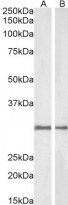
ARG63196 anti-NQO1 antibody WB image
Western blot: 35 µg of Rat (A) and Pig (B) kidney lysates (in RIPA buffer) stained with ARG63196 anti-NQO1 antibody at 1 µg/ml dilution and incubated at RT for 1 hour.


 New Products


