


anti-PDI antibody
Key features and details
- 产品描述:
- 反应物种:
- 应用:
- 特异性:
- 宿主:
- 克隆:
- 靶点名称:
- 抗原物种:
- 抗原:
-
Brand:

 Product Details
| 产品描述 | Rabbit Polyclonal antibody recognizes PDI |
|---|---|
| 反应物种 | Hu, Ms, Rat, Bov, Dog, Gpig, Hm, Mk, Pig, Sheep, Xenopus laevis |
| 应用 | ICC/IF, IHC, IP, WB |
| 特异性 | Detects ~58kDa. |
| 宿主 | Rabbit |
| 克隆 | Polyclonal |
| 靶点名称 | PDI |
| 抗原物种 | Rat |
| 抗原 | KLH-conjugated synthetic peptide from Rat PDI |
| 偶联标记 | Un-conjugated |
| 別名 | PDA2; Pancreas-specific protein disulfide isomerase; PDIR; PDIP; PDI; Protein disulfide-isomerase A2; PDIp; EC 5.3.4.1 |
| 应用建议 |
| ||||||||||
|---|---|---|---|---|---|---|---|---|---|---|---|
| 应用说明 | * The dilutions indicate recommended starting dilutions and the optimal dilutions or concentrations should be determined by the scientist. |
| 形式 | Liquid |
|---|---|
| 纯化 | Affinity purification with immunogen. |
| 缓冲液 | PBS (pH 7.4), 0.09% Sodium azide and 50% Glycerol |
| 抗菌剂 | 0.09% Sodium azide |
| 稳定剂 | 50% Glycerol |
| 浓度 | 1 mg/ml |
| 存放说明 | For continuous use, store undiluted antibody at 2-8°C for up to a week. For long-term storage, aliquot and store at -20°C. Storage in frost free freezers is not recommended. Avoid repeated freeze/thaw cycles. Suggest spin the vial prior to opening. The antibody solution should be gently mixed before use. |
| 注意事项 | For laboratory research only, not for drug, diagnostic or other use. |
| 数据库连接 | |
|---|---|
| 基因名称 | Pdia2 |
| 全名 | protein disulfide isomerase family A, member 2 |
| 背景介绍 | Protein disulfide isomerases (EC 5.3.4.1), such as PDIP, are endoplasmic reticulum (ER) resident proteins that catalyze protein folding and thiol-disulfide interchange reactions (Desilva et al., 1996 [PubMed 8561901]).[supplied by OMIM, Mar 2008] |
| 生物功能 | Acts as an intracellular estrogen-binding protein. May be involved in modulating cellular levels and biological functions of estrogens in the pancreas. May act as a chaperone that inhibits aggregation of misfolded proteins. [UniProt] |
| 细胞定位 | Endoplasmic Reticulum, Endoplasmic reticulum lumen |
| 预测分子量 | 58 kDa |
| 翻译后修饰 | The disulfide-linked homodimer exhibits an enhanced chaperone activity. Glycosylated. |
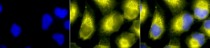
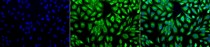
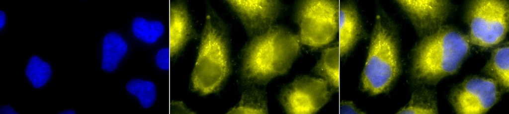
ARG22292 anti-PDI antibody ICC/IF image
Immunocytochemistry: 2% Formaldehyde (20 min at RT) fixed HeLa cells stained with ARG22292 anti-PDI antibody (yellow) at 1:100 dilution (12 hours at 4°C). Counterstain: DAPI (blue) nuclear stain at 1:40000 for 120 min at RT. Magnification: 100x. Left: DAPI (blue) nuclear stain, Middle: Primary antibody, Right: Composite.

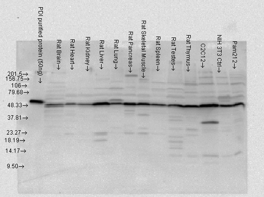
ARG22292 anti-PDI antibody WB image
Western blot: 50 ng of PDI purified protein and 15 µg of Rat tissue/cell lysates stained with ARG22292 anti-PDI antibody at 1:4000 dilution.
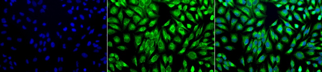
ARG22292 anti-PDI antibody ICC/IF image
Immunocytochemistry: 2% Formaldehyde (20 min at RT) fixed HeLa cells stained with ARG22292 anti-PDI antibody (green) at 1:100 dilution (12 hours at 4°C). Counterstain: DAPI (blue) nuclear stain at 1:40000 for 120 min at RT. Magnification: 20x. Left: DAPI (blue) nuclear stain, Middle: Primary antibody, Right: Composite.


 New Products


