


anti-RP2 / XRP2 antibody


Key features and details
- 产品描述:
- 反应物种:
- 应用:
- 宿主:
- 克隆:
- 同位型:
- 靶点名称:
- 抗原物种:
- 抗原:
-
Brand:

 Product Details
| 产品描述 | Rabbit Polyclonal antibody recognizes RP2 / XRP2 |
|---|---|
| 反应物种 | Hu, Ms, Rat |
| 应用 | FACS, IHC-P, WB |
| 宿主 | Rabbit |
| 克隆 | Polyclonal |
| 同位型 | IgG |
| 靶点名称 | RP2 / XRP2 |
| 抗原物种 | Human |
| 抗原 | Recombinant protein corresponding to D244-M348 of Human RP2 / XRP2. |
| 偶联标记 | Un-conjugated |
| 別名 | TBCCD2; Protein XRP2; XRP2; DELXp11.3; NM23-H10; NME10 |
| 应用建议 |
| ||||||||
|---|---|---|---|---|---|---|---|---|---|
| 应用说明 | IHC-P: Antigen Retrieval: Heat mediation was performed in Citrate buffer (pH 6.0) for 20 min. * The dilutions indicate recommended starting dilutions and the optimal dilutions or concentrations should be determined by the scientist. | ||||||||
| 实际分子量 | ~ 39 kDa |
| 形式 | Liquid |
|---|---|
| 纯化 | Affinity purification with immunogen. |
| 缓冲液 | 0.2% Na2HPO4, 0.9% NaCl, 0.05% Sodium azide and 4% Trehalose. |
| 抗菌剂 | 0.05% Sodium azide |
| 稳定剂 | 4% Trehalose |
| 浓度 | 0.5 mg/ml |
| 存放说明 | For continuous use, store undiluted antibody at 2-8°C for up to a week. For long-term storage, aliquot and store at -20°C or below. Storage in frost free freezers is not recommended. Avoid repeated freeze/thaw cycles. Suggest spin the vial prior to opening. The antibody solution should be gently mixed before use. |
| 注意事项 | For laboratory research only, not for drug, diagnostic or other use. |
| 数据库连接 | |
|---|---|
| 基因名称 | RP2 |
| 全名 | retinitis pigmentosa 2 (X-linked recessive) |
| 背景介绍 | The RP2 locus has been implicated as one cause of X-linked retinitis pigmentosa. The predicted gene product shows homology with human cofactor C, a protein involved in the ultimate step of beta-tubulin folding. Progressive retinal degeneration may therefore be due to the accumulation of incorrectly-folded photoreceptor or neuron-specific tubulin isoforms followed by progressive cell death [provided by RefSeq, Jul 2008] |
| 生物功能 | Acts as a GTPase-activating protein (GAP) involved in trafficking between the Golgi and the ciliary membrane. Involved in localization of proteins, such as NPHP3, to the cilium membrane by inducing hydrolysis of GTP ARL3, leading to the release of UNC119 (or UNC119B). Acts as a GTPase-activating protein (GAP) for tubulin in concert with tubulin-specific chaperone C, but does not enhance tubulin heterodimerization. Acts as guanine nucleotide dissociation inhibitor towards ADP-ribosylation factor-like proteins. [UniProt] |
| 细胞定位 | Cell membrane; Lipid-anchor; Cytoplasmic side. Cell projection, cilium. Note=Detected predominantly at the plasma membrane of rod and cone photoreceptors. Not detected in the nucleus. [UniProt] |
| 预测分子量 | 40 kDa |
| 翻译后修饰 | Myristoylated on Gly-2; which may be required for membrane targeting. Palmitoylated on Cys-3; which may be required for plasma membrane targeting (Probable). Mutation of Cys-3 targets the protein to internal membranes. [UniProt] |

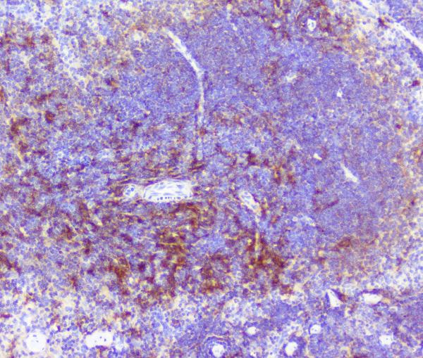
ARG42957 anti-RP2 / XRP2 antibody IHC-P image
Immunohistochemistry: Paraffin-embedded Mouse spleen tissue. Antigen Retrieval: Heat mediation was performed in Citrate buffer (pH 6.0) for 20 min. The tissue section was blocked with 10% goat serum. The tissue section was then stained with ARG42957 anti-RP2 / XRP2 antibody at 1 µg/ml dilution, overnight at 4°C.

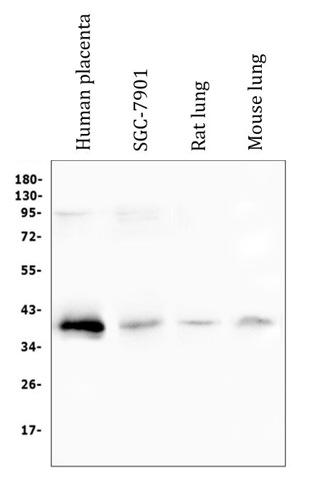
ARG42957 anti-RP2 / XRP2 antibody WB image
Western blot: 50 µg of sample under reducing conditions. Human placenta, SGC-7901, Rat lung and Mouse lung lysates stained with ARG42957 anti-RP2 / XRP2 antibody at 0.5 µg/ml dilution, overnight at 4°C.
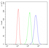
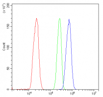
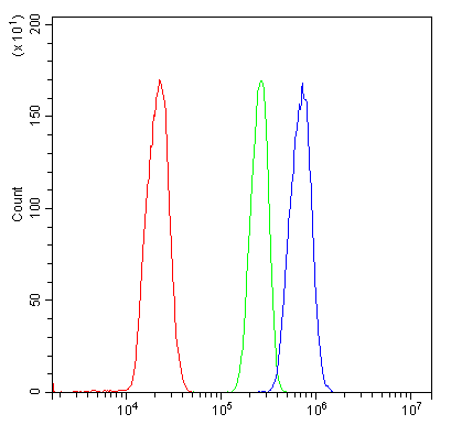
ARG42957 anti-RP2 / XRP2 antibody FACS image
Flow Cytometry: A431 cells were blocked with 10% normal goat serum and then stained with ARG42957 anti-RP2 / XRP2 antibody (blue) at 1 µg/10^6 cells for 30 min at 20°C, followed by incubation with DyLight®488 labelled secondary antibody. Isotype control antibody (green) was Rabbit IgG (1 µg/10^6 cells) used under the same conditions. Unlabelled sample (red) was also used as a control.

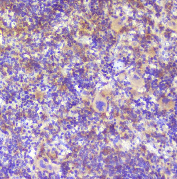
ARG42957 anti-RP2 / XRP2 antibody IHC-P image
Immunohistochemistry: Paraffin-embedded Rat spleen tissue. Antigen Retrieval: Heat mediation was performed in Citrate buffer (pH 6.0) for 20 min. The tissue section was blocked with 10% goat serum. The tissue section was then stained with ARG42957 anti-RP2 / XRP2 antibody at 1 µg/ml dilution, overnight at 4°C.
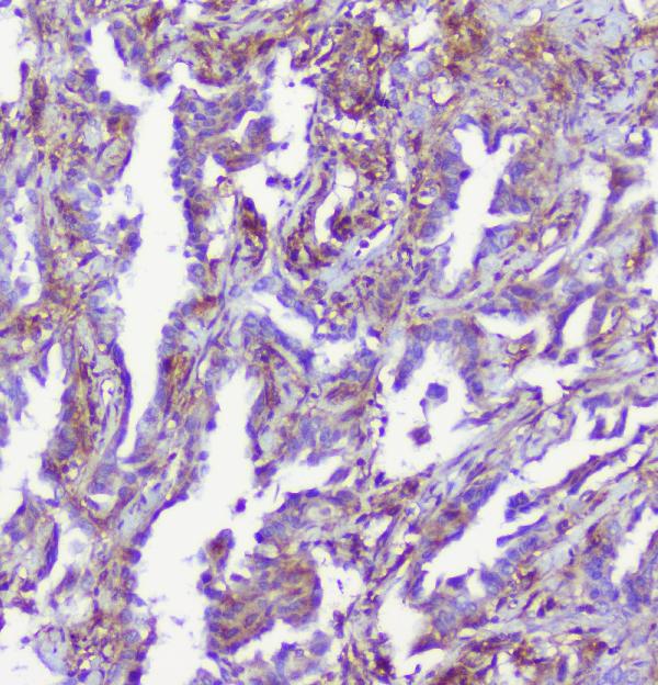
ARG42957 anti-RP2 / XRP2 antibody IHC-P image
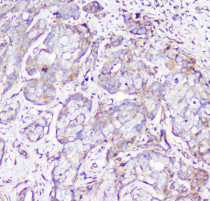
Immunohistochemistry: Paraffin-embedded Human mammary cancer tissue. Antigen Retrieval: Heat mediation was performed in Citrate buffer (pH 6.0) for 20 min. The tissue section was blocked with 10% goat serum. The tissue section was then stained with ARG42957 anti-RP2 / XRP2 antibody at 1 µg/ml dilution, overnight at 4°C.
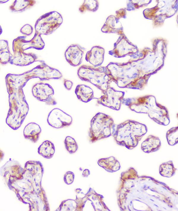
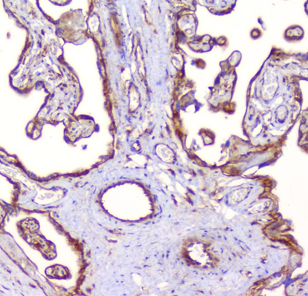
ARG42957 anti-RP2 / XRP2 antibody IHC-P image
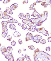
Immunohistochemistry: Paraffin-embedded Human placenta tissue. Antigen Retrieval: Heat mediation was performed in Citrate buffer (pH 6.0) for 20 min. The tissue section was blocked with 10% goat serum. The tissue section was then stained with ARG42957 anti-RP2 / XRP2 antibody at 1 µg/ml dilution, overnight at 4°C.
ARG42957 anti-RP2 / XRP2 antibody IHC-P image
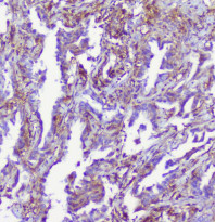
Immunohistochemistry: Paraffin-embedded Human lung cancer tissue. Antigen Retrieval: Heat mediation was performed in Citrate buffer (pH 6.0) for 20 min. The tissue section was blocked with 10% goat serum. The tissue section was then stained with ARG42957 anti-RP2 / XRP2 antibody at 1 µg/ml dilution, overnight at 4°C.

ARG42957 anti-RP2 / XRP2 antibody IHC-P image
Immunohistochemistry: Paraffin-embedded Human placenta tissue. Antigen Retrieval: Heat mediation was performed in Citrate buffer (pH 6.0) for 20 min. The tissue section was blocked with 10% goat serum. The tissue section was then stained with ARG42957 anti-RP2 / XRP2 antibody at 1 µg/ml dilution, overnight at 4°C.
ARG42957 anti-RP2 / XRP2 antibody FACS image
Flow Cytometry: THP-1 cells were blocked with 10% normal goat serum and then stained with ARG42957 anti-RP2 / XRP2 antibody (blue) at 1 µg/10^6 cells for 30 min at 20°C, followed by incubation with DyLight®488 labelled secondary antibody. Isotype control antibody (green) was Rabbit IgG (1 µg/10^6 cells) used under the same conditions. Unlabelled sample (red) was also used as a control.


 New Products


