


anti-RPL19 antibody


Key features and details
- 产品描述:
- 反应物种:
- 预测物种:
- 应用:
- 宿主:
- 克隆:
- 同位型:
- 靶点名称:
- 抗原物种:
-
Brand:

 Product Details
| 产品描述 | Rabbit Polyclonal antibody recognizes RPL19 |
|---|---|
| 反应物种 | Hu, Ms, Rat |
| 预测物种 | Bov, Chk, Dog, Mk, Rb |
| 应用 | FACS, ICC/IF, WB |
| 宿主 | Rabbit |
| 克隆 | Polyclonal |
| 同位型 | IgG |
| 靶点名称 | RPL19 |
| 抗原物种 | Human |
| 抗原 | Synthetic peptide corresponding to aa. 132-170 of Human RPL19. (FKNKRILMEHIHKLKADKARKKLLADQAEARRSKTKEAR) |
| 偶联标记 | Un-conjugated |
| 別名 | 60S ribosomal protein L19; L19 |
| 应用建议 |
| ||||||||
|---|---|---|---|---|---|---|---|---|---|
| 应用说明 | * The dilutions indicate recommended starting dilutions and the optimal dilutions or concentrations should be determined by the scientist. |
| 形式 | Liquid |
|---|---|
| 纯化 | Affinity purification with immunogen. |
| 缓冲液 | 0.2% Na2HPO4, 0.9% NaCl, 0.05% Sodium azide and 5% BSA. |
| 抗菌剂 | 0.05% Sodium azide |
| 稳定剂 | 5% BSA |
| 浓度 | 0.5 mg/ml |
| 存放说明 | For continuous use, store undiluted antibody at 2-8°C for up to a week. For long-term storage, aliquot and store at -20°C or below. Storage in frost free freezers is not recommended. Avoid repeated freeze/thaw cycles. Suggest spin the vial prior to opening. The antibody solution should be gently mixed before use. |
| 注意事项 | For laboratory research only, not for drug, diagnostic or other use. |
| 数据库连接 | |
|---|---|
| 基因名称 | RPL19 |
| 全名 | ribosomal protein L19 |
| 背景介绍 | Ribosomes, the organelles that catalyze protein synthesis, consist of a small 40S subunit and a large 60S subunit. Together these subunits are composed of 4 RNA species and approximately 80 structurally distinct proteins. This gene encodes a ribosomal protein that is a component of the 60S subunit. The protein belongs to the L19E family of ribosomal proteins. It is located in the cytoplasm. As is typical for genes encoding ribosomal proteins, there are multiple processed pseudogenes of this gene dispersed through the genome. [provided by RefSeq, Jul 2008] |
| 预测分子量 | 23 kDa |
| 翻译后修饰 | Citrullinated by PADI4. [UniProt] |
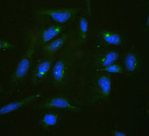
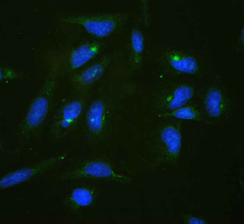
ARG40701 anti-RPL19 antibody ICC/IF image
Immunofluorescence: U20S cells stained with ARG40701 anti-RPL19 antibody at 2 µg/ml, overnight at 4°C. Cells was counterstained with DAPI (blue).
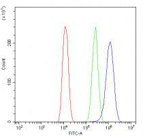
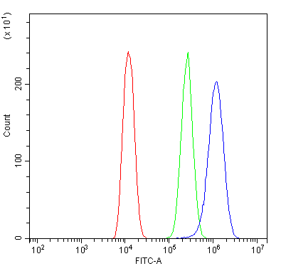
ARG40701 anti-RPL19 antibody FACS image
Flow Cytometry: U20S cells were blocked with 10% normal goat serum and stained with ARG40701 anti-RPL19 antibody (blue) at 1 µg/10^6 cells for 30 min at 20°C, followed by incubation with DyLight®488 labelled secondary antibody. Isotype control antibody (green) was rabbit IgG (1 µg/10^6 cells) used under the same conditions. Unlabelled sample (red) was also used as a control.

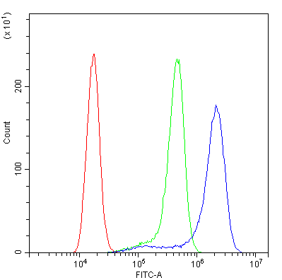
ARG40701 anti-RPL19 antibody FACS image
Flow Cytometry: A431 cells were blocked with 10% normal goat serum and stained with ARG40701 anti-RPL19 antibody (blue) at 1 µg/10^6 cells for 30 min at 20°C, followed by incubation with DyLight®488 labelled secondary antibody. Isotype control antibody (green) was rabbit IgG (1 µg/10^6 cells) used under the same conditions. Unlabelled sample (red) was also used as a control.
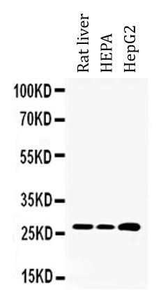
ARG40701 anti-RPL19 antibody WB image
Western blot: Rat liver, HEPA and HepG2 whole cell lysates stained with ARG40701 anti-RPL19 antibody at 0.5 µg/ml.

 New Products


