


anti-TAK1 / MAP3K7 antibody


Key features and details
- 产品描述:
- 反应物种:
- 预测物种:
- 应用:
- 特异性:
- 宿主:
- 克隆:
- 同位型:
- 靶点名称:
-
Brand:

 Product Details
| 产品描述 | Goat Polyclonal antibody recognizes TAK1 / MAP3K7 |
|---|---|
| 反应物种 | Hu, Ms |
| 预测物种 | Cow, Rat |
| 应用 | FACS, WB |
| 特异性 | This antibody is expected to recognise isoform A (NP_003179.1) and isoform B (NP_663304.1). |
| 宿主 | Goat |
| 克隆 | Polyclonal |
| 同位型 | IgG |
| 靶点名称 | TAK1 / MAP3K7 |
| 抗原物种 | Human |
| 抗原 | C-AELDQDEKDQQNT |
| 偶联标记 | Un-conjugated |
| 別名 | TAK1; Mitogen-activated protein kinase kinase kinase 7; EC 2.7.11.25; Transforming growth factor-beta-activated kinase 1; TGF-beta-activated kinase 1; MEKK7; TGF1a |
| 应用建议 |
| ||||||
|---|---|---|---|---|---|---|---|
| 应用说明 | WB: Recommend incubate at RT for 1h. * The dilutions indicate recommended starting dilutions and the optimal dilutions or concentrations should be determined by the scientist. |
| 形式 | Liquid |
|---|---|
| 纯化 | Purified from goat serum by ammonium sulphate precipitation followed by antigen affinity chromatography using the immunizing peptide. |
| 缓冲液 | Tris saline (pH 7.3), 0.02% Sodium azide and 0.5% BSA |
| 抗菌剂 | 0.02% Sodium azide |
| 稳定剂 | 0.5% BSA |
| 浓度 | 0.5 mg/ml |
| 存放说明 | For continuous use, store undiluted antibody at 2-8°C for up to a week. For long-term storage, aliquot and store at -20°C or below. Storage in frost free freezers is not recommended. Avoid repeated freeze/thaw cycles. Suggest spin the vial prior to opening. The antibody solution should be gently mixed before use. |
| 注意事项 | For laboratory research only, not for drug, diagnostic or other use. |
| 数据库连接 | Swiss-port # O43318 Human Mitogen-activated protein kinase kinase kinase 7 Swiss-port # Q62073 Mouse Mitogen-activated protein kinase kinase kinase 7 |
|---|---|
| 背景介绍 | The protein encoded by this gene is a member of the serine/threonine protein kinase family. This kinase mediates the signaling transduction induced by TGF beta and morphogenetic protein (BMP), and controls a variety of cell functions including transcription regulation and apoptosis. In response to IL-1, this protein forms a kinase complex including TRAF6, MAP3K7P1/TAB1 and MAP3K7P2/TAB2; this complex is required for the activation of nuclear factor kappa B. This kinase can also activate MAPK8/JNK, MAP2K4/MKK4, and thus plays a role in the cell response to environmental stresses. Four alternatively spliced transcript variants encoding distinct isoforms have been reported. [provided by RefSeq, Jul 2008] |
| 研究领域 | Cancer antibody; Cell Biology and Cellular Response antibody; Immune System antibody; Signaling Transduction antibody |
| 预测分子量 | 67 kDa |
| 翻译后修饰 | Association with TAB1/MAP3K7IP1 promotes autophosphorylation at Ser-192 and subsequent activation. Association with TAB2/MAP3K7IP2, itself associated with free unanchored Lys-63 polyubiquitin chain, promotes autophosphorylation and subsequent activation of MAP3K7. Dephosphorylation at Ser-192 by PPM1B/PP2CB and at Thr-187 by PP2A and PPP6C leads to inactivation. 'Lys-48'-linked polyubiquitination at Lys-72 is induced by TNFalpha, and leads to proteasomal degradation. Undergoes 'Lys-48'-linked polyubiquitination catalyzed by ITCH (By similarity). Requires 'Lys-63'-linked polyubiquitination for autophosphorylation and subsequent activation. 'Lys-63'-linked ubiquitination does not lead to proteasomal degradation. Deubiquitinated by CYLD, a protease that selectively cleaves 'Lys-63'-linked ubiquitin chains. Deubiquitinated by Y.enterocolitica YopP. |
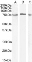
ARG63986 anti-TAK1 / MAP3K7 antibody WB image
Western blot: 35 µg of HeLa (A), U937 (B) and Daudi (C) cell lysates (in RIPA buffer) stained with ARG63986 anti-TAK1 / MAP3K7 antibody at 1 µg/ml (A, B) and 2 µg/ml (C) dilutions and incubated at RT for 1 hour.
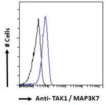
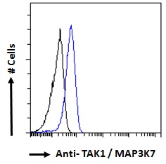
ARG63986 anti-TAK1 / MAP3K7 antibody FACS image
Flow Cytometry: Paraformaldehyde-fixed HeLa cells permeabilized with 0.5% Triton. Cells were stained with ARG63986 anti-TAK1 / MAP3K7 antibody (blue line) at 10 µg/ml dilution for 1 hour, followed by incubation with Alexa FluorR 488 labelled secondary antibody. IgG control: Unimmunized goat IgG (black line).
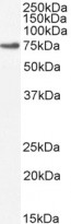
ARG63986 anti-TAK1 / MAP3K7 antibody WB image
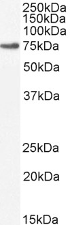
Western blot: 35 µg of NIH/3T3 cell lysate (in RIPA buffer) stained with ARG63986 anti-TAK1 / MAP3K7 antibody at 2 µg/ml dilution and incubated at RT for 1 hour.


 New Products


