


anti-TGF beta Receptor 1 antibody


Key features and details
- 产品描述:
- 反应物种:
- 预测物种:
- 应用:
- 特异性:
- 宿主:
- 克隆:
- 靶点名称:
- 抗原物种:
-
Brand:

 Product Details
| 产品描述 | Goat Polyclonal antibody recognizes TGF beta Receptor 1 |
|---|---|
| 反应物种 | Hu, Rat |
| 预测物种 | Ms, Cow, Dog, Pig |
| 应用 | FACS, IHC-P, WB |
| 特异性 | This antibody is expected to recognize both reported isoforms (NP_004603.1; NP_001124388.1). The immunizing peptide represents part of a potential extracellular domain. |
| 宿主 | Goat |
| 克隆 | Polyclonal |
| 靶点名称 | TGF beta Receptor 1 |
| 抗原物种 | Human |
| 抗原 | Synthetic peptide around the internal region of Human TGF beta Receptor 1 (TDGLCFVSVTETTD) |
| 偶联标记 | Un-conjugated |
| 別名 | ESS1; TGF-beta receptor type-1; ALK5; LDS2A; AAT5; Activin receptor-like kinase 5; LDS1; tbetaR-I; Activin A receptor type II-like protein kinase of 53kD; MSSE; ALK-5; ACVRLK4; TGFR-1; TGF-beta receptor type I; TbetaR-I; LDS1A; Serine/threonine-protein kinase receptor R4; EC 2.7.11.30; Transforming growth factor-beta receptor type I; SKR4; TGF-beta type I receptor |
| 应用建议 |
| ||||||||
|---|---|---|---|---|---|---|---|---|---|
| 应用说明 | WB: Recommend incubate at RT for 1h. IHC-P: Antigen Retrieval: Steam tissue section in Citrate buffer (pH 6.0). * The dilutions indicate recommended starting dilutions and the optimal dilutions or concentrations should be determined by the scientist. |
| 形式 | Liquid |
|---|---|
| 纯化 | Affinity purification with immunogen. |
| 缓冲液 | Tris saline (pH 7.3), 0.02% Sodium azide and 0.5% BSA |
| 抗菌剂 | 0.02% Sodium azide |
| 稳定剂 | 0.5% BSA |
| 浓度 | 0.5 mg/ml |
| 存放说明 | For continuous use, store undiluted antibody at 2-8°C for up to a week. For long-term storage, aliquot and store at -20°C or below. Storage in frost free freezers is not recommended. Avoid repeated freeze/thaw cycles. Suggest spin the vial prior to opening. The antibody solution should be gently mixed before use. |
| 注意事项 | For laboratory research only, not for drug, diagnostic or other use. |
| 数据库连接 | |
|---|---|
| 基因名称 | TGFBR1 |
| 全名 | transforming growth factor, beta receptor 1 |
| 背景介绍 | The protein encoded by this gene forms a heteromeric complex with type II TGF-beta receptors when bound to TGF-beta, transducing the TGF-beta signal from the cell surface to the cytoplasm. The encoded protein is a serine/threonine protein kinase. Mutations in this gene have been associated with Loeys-Dietz aortic aneurysm syndrome (LDAS). Multiple transcript variants encoding different isoforms have been found for this gene. [provided by RefSeq, Aug 2008] |
| 生物功能 | Transmembrane serine/threonine kinase forming with the TGF-beta type II serine/threonine kinase receptor, TGFBR2, the non-promiscuous receptor for the TGF-beta cytokines TGFB1, TGFB2 and TGFB3. Transduces the TGFB1, TGFB2 and TGFB3 signal from the cell surface to the cytoplasm and is thus regulating a plethora of physiological and pathological processes including cell cycle arrest in epithelial and hematopoietic cells, control of mesenchymal cell proliferation and differentiation, wound healing, extracellular matrix production, immunosuppression and carcinogenesis. The formation of the receptor complex composed of 2 TGFBR1 and 2 TGFBR2 molecules symmetrically bound to the cytokine dimer results in the phosphorylation and the activation of TGFBR1 by the constitutively active TGFBR2. Activated TGFBR1 phosphorylates SMAD2 which dissociates from the receptor and interacts with SMAD4. The SMAD2-SMAD4 complex is subsequently translocated to the nucleus where it modulates the transcription of the TGF-beta-regulated genes. This constitutes the canonical SMAD-dependent TGF-beta signaling cascade. Also involved in non-canonical, SMAD-independent TGF-beta signaling pathways. For instance, TGFBR1 induces TRAF6 autoubiquitination which in turn results in MAP3K7 ubiquitination and activation to trigger apoptosis. Also regulates epithelial to mesenchymal transition through a SMAD-independent signaling pathway through PARD6A phosphorylation and activation. [UniProt] |
| 研究领域 | Cancer antibody; Cell Biology and Cellular Response antibody; Developmental Biology antibody; Metabolism antibody; Signaling Transduction antibody |
| 预测分子量 | 55.6 kDa (NP_036907.2) |
| 翻译后修饰 | Phosphorylated at basal levels in the absence of ligand. Activated upon phosphorylation by TGFBR2, mainly in the GS domain. Phosphorylation in the GS domain abrogates FKBP1A-binding. N-Glycosylated. Ubiquitinated; undergoes ubiquitination catalyzed by several E3 ubiquitin ligases including SMURF1, SMURF2 and NEDD4L2. Results in the proteasomal and/or lysosomal degradation of the receptor thereby negatively regulating its activity. Deubiquitinated by USP15, leading to stabilization of the protein and enhanced TGF-beta signal. Its ubiquitination and proteasome-mediated degradation is negatively regulated by SDCBP (PubMed:25893292). |

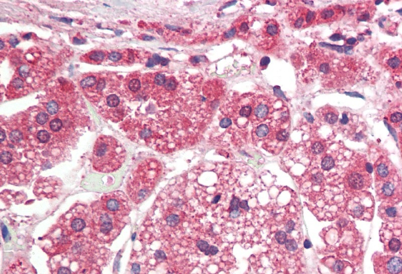
ARG65658 anti-TGF beta Receptor 1 antibody IHC-P image
Immunohistochemistry: Paraffin-embedded Human adrenal gland tissue. Antigen Retrieval: Steam tissue section in Citrate buffer (pH 6.0). The tissue section was stained with ARG65658 anti-TGF beta Receptor 1 antibody at 5 µg/ml dilution followed by AP-staining.

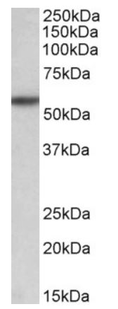
ARG65658 anti-TGF beta Receptor 1 antibody WB image
Western blot: 35 μg of Rat liver lysate stained with ARG65658 anti-TGF beta Receptor 1 antibody at 0.1 μg/ml dilution and incubated at RT for 1 hour.
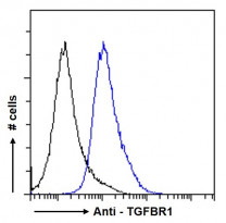
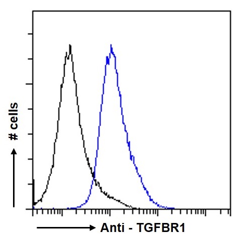
ARG65658 anti-TGF beta Receptor 1 antibody FACS image
Flow Cytometry: Paraformaldehyde-fixed HeLa cells permeabilized with 0.5% Triton. Cells were stained with ARG65658 anti-TGF beta Receptor 1 antibody (blue line) at 10 µg/ml dilution for 1 hour, followed by incubation with Alexa Fluor® 488 labelled secondary antibody. IgG control: Unimmunized goat IgG (black line), followed by incubation with Alexa Fluor® 488 labelled secondary antibody.
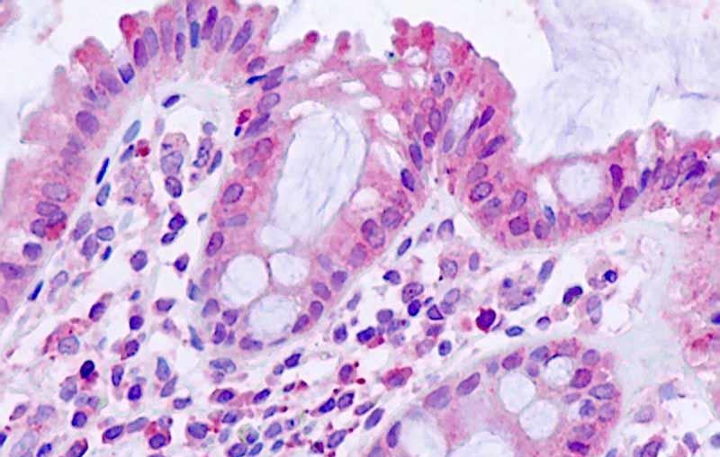
ARG65658 anti-TGF beta Receptor 1 antibody IHC-P image
Immunohistochemistry: Paraffin-embedded Human small intestine tissue. Antigen Retrieval: Steam tissue section in Citrate buffer (pH 6.0). The tissue section was stained with ARG65658 anti-TGF beta Receptor 1 antibody at 5 µg/ml dilution followed by AP-staining.


 New Products


