


anti-ZO1 antibody


Key features and details
- 产品描述:
- 反应物种:
- 应用:
- 宿主:
- 克隆:
- 同位型:
- 靶点名称:
- 抗原物种:
- 抗原:
-
Brand:

CAT.NO. : ARG42916
US$ Please choose
US$ Please choose
Size:
Trail, Bulk size or Custom requests Please contact us
*产品价格可能会有所调整,请以品牌方官网实时更新的价格为准,以确保准确性。
 Product Details
概述
| 产品描述 | Rabbit Polyclonal antibody recognizes ZO1 |
|---|---|
| 反应物种 | Hu, Ms |
| 应用 | ICC/IF, IHC-P, WB |
| 宿主 | Rabbit |
| 克隆 | Polyclonal |
| 同位型 | IgG |
| 靶点名称 | ZO1 |
| 抗原物种 | Human |
| 抗原 | Human ZO1. |
| 偶联标记 | Un-conjugated |
| 別名 | Zonula occludens protein 1; Tight junction protein ZO-1; Tight junction protein 1; Zona occludens protein 1; ZO-1 |
应用说明
| 应用建议 |
| ||||||||
|---|---|---|---|---|---|---|---|---|---|
| 应用说明 | * The dilutions indicate recommended starting dilutions and the optimal dilutions or concentrations should be determined by the scientist. |
属性
| 形式 | Liquid |
|---|---|
| 纯化 | Affinity purified. |
| 缓冲液 | 100 mM Tris Glycine (pH 7.0), 0.025% ProClin 300, 20% Glycerol and 1% BSA. |
| 抗菌剂 | 0.025% ProClin 300 |
| 稳定剂 | 20% Glycerol and 1% BSA |
| 浓度 | 0.47 mg/ml |
| 存放说明 | For continuous use, store undiluted antibody at 2-8°C for up to a week. For long-term storage, aliquot and store at -20°C. Storage in frost free freezers is not recommended. Avoid repeated freeze/thaw cycles. Suggest spin the vial prior to opening. The antibody solution should be gently mixed before use. |
| 注意事项 | For laboratory research only, not for drug, diagnostic or other use. |
生物信息
| 数据库连接 | |
|---|---|
| 基因名称 | TJP1 |
| 全名 | tight junction protein 1 |
| 背景介绍 | This gene encodes a member of the membrane-associated guanylate kinase (MAGUK) family of proteins, and acts as a tight junction adaptor protein that also regulates adherens junctions. Tight junctions regulate the movement of ions and macromolecules between endothelial and epithelial cells. The multidomain structure of this scaffold protein, including a postsynaptic density 95/disc-large/zona occludens (PDZ) domain, a Src homology (SH3) domain, a guanylate kinase (GuK) domain and unique (U) motifs all help to co-ordinate binding of transmembrane proteins, cytosolic proteins, and F-actin, which are required for tight junction function. Alternative splicing results in multiple transcript variants encoding different isoforms. [provided by RefSeq, Aug 2017] |
| 生物功能 | TJP1, TJP2, and TJP3 are closely related scaffolding proteins that link tight junction (TJ) transmembrane proteins such as claudins, junctional adhesion molecules, and occludin to the actin cytoskeleton (PubMed:7798316, PubMed:9792688). The tight junction acts to limit movement of substances through the paracellular space and as a boundary between the compositionally distinct apical and basolateral plasma membrane domains of epithelial and endothelial cells. Necessary for lumenogenesis, and particularly efficient epithelial polarization and barrier formation (By similarity). Plays a role in the regulation of cell migration by targeting CDC42BPB to the leading edge of migrating cells (PubMed:21240187). Plays an important role in podosome formation and associated function, thus regulating cell adhesion and matrix remodeling (PubMed:20930113). With TJP2 and TJP3, participates to the junctional retention and stability of the transcription factor DBPA, but is not involved in its shuttling to the nucleus (By similarity). [UniProt] |
| 细胞定位 | Cell membrane; Peripheral membrane protein; Cytoplasmic side. Cell junction, tight junction. Cell junction. Cell junction, gap junction. Cell projection, podosome. Note=Moves from the cytoplasm to the cell membrane concurrently with cell-cell contact (PubMed:7798316). At podosomal sites, is predominantly localized in the ring structure surrounding the actin core (PubMed:20930113). [UniProt] |
| 预测分子量 | 195 kDa |
| 翻译后修饰 | Phosphorylated. Dephosphorylated by PTPRJ. [UniProt] |
检测图片 (4)
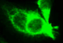
ARG42916 anti-ZO1 antibody ICC/IF image
Immunofluorescence: HT-29 cells were fixed with 4% paraformaldehyde for 10 min at RT, permeabilized with 0.1% NP-40 for 10 min at RT then blocked with 5% BSA for 30 min at room temperature. Cells were stained with ARG42916 anti-ZO1 antibody at 1:150 dilution and 4°C.
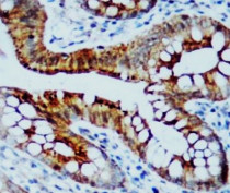
ARG42916 anti-ZO1 antibody IHC-P image
Immunohistochemistry: Paraffin-embedded Human colorectal carcinoma tissue stained with ARG42916 anti-ZO1 antibody at 1:100 dilution.

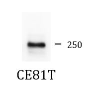
ARG42916 anti-ZO1 antibody WB image
Western blot: 50 µg of CE81T cell lysate stained with ARG42916 anti-ZO1 antibody at 1:500 dilution.
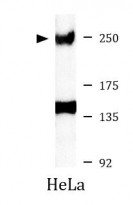
ARG42916 anti-ZO1 antibody WB image
Western blot: 30 µg of HeLa cell lysate stained with ARG42916 anti-ZO1 antibody at 1:500 dilution.


 New Products


