Claudin 2 Rabbit Polyclonal Antibody

Key features and details
- Reactivity:Human, Mouse, Rat, Bovine, Dog, Pig
- Application:WB, IHC, ICC/IF
- Host:Rabbit
- Clonality:polyclonal
- Target:Claudin 2
-
Brand:

CAT.NO. : ARA6424
RMB Please choose
RMB Please choose
*产品价格可能会有所调整,请以品牌方官网实时更新的价格为准,以确保准确性。
 Product Details
Background
Claudin-2 is a protein that in humans is encoded by the CLDN2 gene. It belongs to the group of claudins. Members of the claudin protein family, such as CLDN2, are expressed in an organ-specific manner and regulate the tissue-specific physiologic properties of tight junctions. Claudin-2 is expressed in cation-leaky epithelia such as that of the kidney proximal tubule. Mice that are deficient in claudin-2 have reduced reabsorption of Na+ in the proximal tubule, consistent with a role in paracellular transport. Similar results have been obtained with cultured cells, as overexpression in claudin-2 lacking cells leads to increase of permeability for small cations. Furthermore, claudin-2 has been shown to form paracellular channels for water.
Application
|
Application |
Dilution Ratio |
|
WB |
1:500 - 1:1000 |
|
IHC |
1:100-1:200 |
|
ICC/IF |
1:100 - 1:500 |
Overview
|
Product Description |
Rabbit polyclonal antibody to Claudin 2 |
|
Immunogen |
KLH-conjugated synthetic peptide encompassing a sequence within the C-term region of human Claudin 2. The exact sequence is proprietary. |
|
Purification Method |
The antibody was purified by immunogen affinity chromatography. |
|
Clonality |
Polyclonal |
|
Form |
Liquid in 0.42% Potassium phosphate, 0.87% Sodium chloride, pH 7.3, 30% glycerol, and 0.01% sodium azide. |
|
Recommended Dilution |
WB (1/500 - 1/1000), IH (1/100 - 1/200), IF/IC (1/100 - 1/500) |
|
Gene Symbol |
CLDN2 |
|
Alternative Names |
Claudin-2; SP82 |
|
Gene ID (Human) |
9075 |
|
Gene ID (Mouse) |
12738 |
|
Protein ID (Human) |
P57739 |
|
Protein ID (Mouse) |
O88552 |
Data

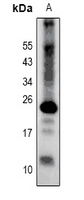
Western blot analysis of Claudin 2 expression in AML12 (A) whole cell lysates. (Predicted band size: 24 kD; Observed band size: 24 kD)
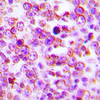
Immunohistochemical analysis of Claudin 2 staining in human tonsil formalin fixed paraffin embedded tissue section. The section was pre-treated using heat mediated antigen retrieval with sodium citrate buffer (pH 6.0). The section was then incubated with the antibody at room temperature and detected using an HRP conjugated compact polymer system. DAB was used as the chromogen. The section was then counterstained with haematoxylin and mounted with DPX.

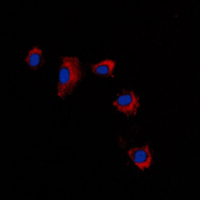
Immunofluorescent analysis of Claudin 2 staining in A431 cells. Formalin-fixed cells were permeabilized with 0.1% Triton X-100 in TBS for 5-10 minutes and blocked with 3% BSA-PBS for 30 minutes at room temperature. Cells were probed with the primary antibody in 3% BSA-PBS and incubated overnight at 4 °C in a humidified chamber. Cells were washed with PBST and incubated with a DyLight 594-conjugated secondary antibody (red) in PBS at room temperature in the dark. DAPI was used to stain the cell nuclei (blue).
Storage
Store at 4°C short term. For long term storage, store at -20°C, avoiding freeze/thaw cycles.
Research Use Only
For Research Use Only. Not for use in diagnostic procedures.
New Products