


Desmin Rabbit Polyclonal Antibody


Key features and details
- Reactivity:Human, Rat, Bovine, Chicken, Dog, Pig
- Application:WB, IHC, ICC/IF
- Host:Rabbit
- Clonality:polyclonal
- Target:Desmin
-
Brand:

CAT.NO. : ARA6485
RMB Please choose
RMB Please choose
*产品价格可能会有所调整,请以品牌方官网实时更新的价格为准,以确保准确性。
 Product Details
Background
Desmin is a type III intermediate filament present in normal smooth, skeletal, and cardiac muscle cells. Analysis by light microscopy suggests desmin localizes towards the periphery of Z-lines in striated muscle fibrils. Desmin connects cytoplasmic dense bodies to membranous dense plaques in smooth muscles. Anti-Desmin stains rhabdomyomas, leiomyosarcoma, rhabdomyosarcoma, leiomyomas, and perivascular cells from skin glomus tumours, and is used to identify the myogenic characteristics of tumours. Desmin can also be found in myofibroblasts and desmoid fibromatosis.
Application
|
Application |
Dilution Ratio |
|
WB |
1:500 - 1:2000 |
|
IHC |
1:100 - 1:200 |
|
ICC/IF |
1:100 - 1:500 |
Overview
|
Immunogen |
KLH-conjugated synthetic peptide encompassing a sequence within the C-term region of human Desmin. The exact sequence is proprietary. |
|
Purification Method |
The antibody was purified by immunogen affinity chromatography. |
|
Clonality |
Polyclonal |
|
Product Form |
Liquid in 0.42% Potassium phosphate, 0.87% Sodium chloride, pH 7.3, 30% glycerol, and 0.01% sodium azide. |
|
Gene Name |
DES |
|
Alternative Names |
Desmin |
|
Human Gene ID |
1674 |
|
Rat Gene ID |
64362 |
|
Human Protein ID |
P17661 |
|
Rat Protein ID |
P48675 |
Data
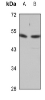
Western blot analysis of Desmin expression in A375 (A), rat heart (B) whole cell lysates. (Predicted band size: 53 kD; Observed band size: 54 kD)

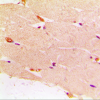
Immunohistochemical analysis of Desmin staining in human muscle formalin fixed paraffin embedded tissue section. The section was pre-treated using heat mediated antigen retrieval with sodium citrate buffer (pH 6.0). The section was then incubated with the antibody at room temperature and detected using an HRP conjugated compact polymer system. DAB was used as the chromogen. The section was then counterstained with haematoxylin and mounted with DPX.
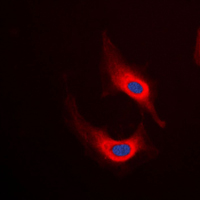
Immunofluorescent analysis of Desmin staining in HeLa cells. Formalin-fixed cells were permeabilized with 0.1% Triton X-100 in TBS for 5-10 minutes and blocked with 3% BSA-PBS for 30 minutes at room temperature. Cells were probed with the primary antibody in 3% BSA-PBS and incubated overnight at 4 °C in a humidified chamber. Cells were washed with PBST and incubated with a DyLight 594-conjugated secondary antibody (red) in PBS at room temperature in the dark. DAPI was used to stain the cell nuclei (blue).
Storage
Store at 4°C short term. For long term storage, store at -20°C, avoiding freeze/thaw cycles.
Research Use Only
For Research Use Only. Not for use in diagnostic procedures.
New Products


