


HMGB2 Rabbit Polyclonal Antibody


Key features and details
- Reactivity:Human, Mouse, Rat
- Application:WB,IHC,IF/ICC
- Host:Rabbit
- Clonality:Polyclonal
- Target:HMGB2
-
Brand:

CAT.NO. : ARA6638
RMB Please choose
RMB Please choose
Size:
Trail, Bulk size or Custom requests Please contact us
*产品价格可能会有所调整,请以品牌方官网实时更新的价格为准,以确保准确性。
 Product Details
Background
HMGB2 encodes a member of the non-histone chromosomal high mobility group protein family. The proteins of this family are chromatin-associated and ubiquitously distributed in the nucleus of higher eukaryotic cells. In vitro studies have demonstrated that this protein is able to efficiently bend DNA and form DNA circles. These studies suggest a role in facilitating cooperative interactions between cis-acting proteins by promoting DNA flexibility. This protein was also reported to be involved in the final ligation step in DNA end-joining processes of DNA double-strand breaks repair and V(D)J recombination.
Application
|
Application |
Dilution Ratio |
|
WB |
1:500 - 1:1000 |
|
IHC |
1:100 - 1:200 |
|
IF/ICC |
1:100 - 1:500 |
Overview
|
Product Description |
Rabbit polyclonal antibody to HMGB2 |
|
Immunogen |
KLH - conjugated synthetic peptide encompassing a sequence within the center region of human HMGB2. The exact sequence is proprietary. |
|
Purification Method |
The antibody was purified by immunogen affinity chromatography |
|
Clonality |
Polyclonal |
|
Product Form |
Liquid in 0.42% Potassium phosphate, 0.87% Sodium chloride, pH 7.3, 30% glycerol, and 0.01% sodium azide |
|
Gene Name |
HMGB2 |
|
Related Names |
HMG2; High mobility group protein B2; High mobility group protein 2; HMG - 2 |
|
Gene ID (Human) |
3148 |
|
Gene ID (Mouse) |
97165 |
|
Protein ID (Human) |
P26583 |
|
Protein ID (Mouse) |
P30681 |
Data

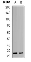
Western blot analysis of HMGB2 expression in Hela (A), K562 (B) whole cell lysates. (Predicted band size: 24 kD; Observed band size: 26 kD)
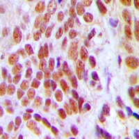
Immunohistochemical analysis of HMGB2 staining in human breast cancer formalin fixed paraffin embedded tissue section. The section was pre-treated using heat mediated antigen retrieval with sodium citrate buffer (pH 6.0). The section was then incubated with the antibody at room temperature and detected using an HRP conjugated compact polymer system. DAB was used as the chromogen. The section was then counterstained with haematoxylin and mounted with DPX.
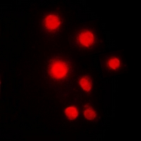
Immunofluorescent analysis of HMGB2 staining in Hela cells. Formalin-fixed cells were permeabilized with 0.1% Triton X-100 in TBS for 5-10 minutes and blocked with 3% BSA-PBS for 30 minutes at room temperature. Cells were probed with the primary antibody in 3% BSA-PBS and incubated overnight at 4 °C in a hidified chamber. Cells were washed with PBST and incubated with a DyLight 594-conjugated secondary antibody (red) in PBS at room temperature in the dark.
Storage
Store at 4°C short term. For long term storage, store at -20°C, avoiding freeze/thaw cycles.
Research Use Only
For Research Use Only. Not for use in diagnostic procedures.
New Products


