


Human CD115/M-CSF R/CSF-1-R ELISA Kit
Key features and details
- Specification:
- Sensitivity:
- Standard Curve Range:
- Standard Curve Gradient:
- Number of Incubations:
- Sample Volume:
- Assay type:
- Operation Duration:
-
Brand:

CAT.NO. : AEHY0168
RMB Please choose
RMB Please choose
Size:
Trail, Bulk size or Custom requests Please contact us
 Product Details
Background
M-CSF receptor, the product of the c-fms proto-oncogene, is a member of the type III subfamily of receptor tyrosine kinases that also includes receptors for SCF and PDGF. These receptors each contain five immunoglobulin-like domains in their extracellular domain (ECD) and a split kinase domain in their intracellular region. M-CSF receptor is expressed primarily on cells of the monocyte/macrophage lineage, dendritic cells, stem cells and in the developing placenta. Human M-CSF receptor cDNA encodes a 972 amino acid (aa) type I membrane protein with a 19 aa signal peptide, a 493 aa extracellular region containing the ligand-binding domain, a 25 aa transmembrane domain and a 435 aa cytoplasmic domain. The human M-CSF R ECD shares 60%, 64%, 72%, 75%, 75% and 76% aa identity with mouse, rat, bovine, canine, feline and equine M-CSF R, respectively. Activators of protein kinase C induce TACE/ADAM17 cleavage of the M-CSF receptor, releasing the functional ligand-binding extracellular domain. M-CSF binding induces receptor homodimerization, resulting in transphosphorylation of specific cytoplasmic tyrosine residues and signal transduction. The intracellular domain of activated M-CSF R binds more than 150 proteins that affect cell proliferation, survival, differentiation and cytoskeletal reorganization. Among these, PI3Kinase, P42/44 ERK and c-Cbl are key transducers of M-CSF R signals. M-CSF R engagement is continuously required for macrophage survival and regulates lineage decisions and maturation of monocytes, macrophages, osteoclasts and DC. M-CSF R and integrin alpha v beta 3 share signaling pathways during osteoclastogenesis and deletion of either causes osteopetrosis. In the brain, microglia expressing increased
M-CSF R are concentrated with Alzheimers a beta peptide, but their role in pathogenesis is unclear.
M-CSF R are concentrated with Alzheimers a beta peptide, but their role in pathogenesis is unclear.
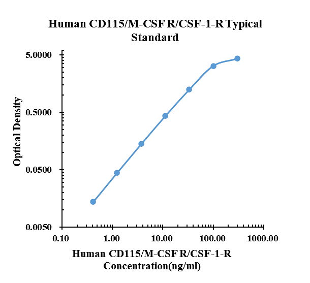
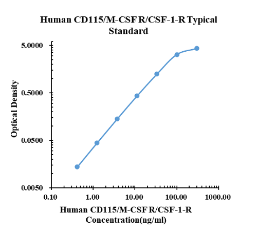
Typical data
|
ng/ml |
O.D. |
Average |
Corrected |
|
|
0.00 |
0.0056 |
0.0054 |
0.0055 |
|
|
0.41 |
0.0188 |
0.0196 |
0.0192 |
0.0137 |
|
1.23 |
0.0493 |
0.0501 |
0.0497 |
0.0442 |
|
3.70 |
0.1445 |
0.1453 |
0.1449 |
0.1394 |
|
11.11 |
0.4360 |
0.4285 |
0.4323 |
0.4268 |
|
33.33 |
1.2330 |
1.2430 |
1.2380 |
1.2325 |
|
100.00 |
3.2110 |
3.1820 |
3.1965 |
3.1910 |
|
300.00 |
4.2906 |
4.2706 |
4.2806 |
4.2751 |
Precision
|
Intra-assay Precision |
Inter-assay Precision |
|||||
|
Sample Number |
S1 |
S2 |
S3 |
S1 |
S2 |
S3 |
|
22 |
22 |
22 |
6 |
6 |
6 |
|
|
Average(ng/ml) |
7.6 |
38.8 |
148.5 |
6.5 |
33.6 |
102.7 |
|
Standard Deviation |
0.3 |
1.8 |
2.7 |
0.4 |
2.0 |
2.5 |
|
Coefficient of Variation(%) |
3.6 |
4.5 |
1.8 |
6.0 |
6.0 |
2.5 |
Inter-assay Precision (Precision between assays) Three samples of known concentration were tested six times on one plate to assess intra-assay precision.
Spike Recovery
The spike recovery was evaluated by spiking 3 levels of human CD115/M-CSF R/CSF-1-R into health human serum sample. The un-spiked serum was used as blank in this experiment.
The recovery ranged from 80% to 99% with an overall mean recovery of 89%.
The recovery ranged from 80% to 99% with an overall mean recovery of 89%.
Sample Values
| Sample Matrix | Sample Evaluated | Range (ng/ml) | Detectable (%) | Mean of Detectable (ng/ml) |
|---|---|---|---|---|
| Serum | 30 | 174.98-741.02 | 100 | 502.94 |
Serum/Plasma – Thirty samples from apparently healthy volunteers were evaluated for the presence of CD115/M-CSF R/CSF-1-R in this assay. No medical histories were available for the donors.
New Products


