


Mitochondria / Caspase dependant Apoptosis Antibody Panel (Caspase3, Caspase9, Cytochrome c, PARP) (WB)

Key features and details
- 产品描述:
- 靶点名称:
- 別名:
-
Brand:

 Product Details
| 货号 | 内含物名称 | 宿主克隆性 | 反应 | 应用 | 包装 |
|---|---|---|---|---|---|
| ARG20006 | anti-Cytochrome C antibody | Rabbit pAb | Hu, Ms, Rat, Bov, Chk, Dog | IP, WB | 25 μg |
| ARG20048 | anti-Caspase 9 (cleaved) antibody | Rabbit pAb | Hu | IP, WB | 25 μg |
| ARG20001 | anti-Caspase 3 antibody [C33] | Mouse mAb | Hu | WB | 25 μg |
| ARG20041 | anti-PARP (cleaved) antibody | Rabbit pAb | Hu | IHC, WB | 25 μg |
| ARG65351 | Goat anti-Rabbit IgG antibody (HRP) | Goat pAb | Rb | ELISA, IHC-P, WB | 50 μl |
| ARG65350 | Goat anti-Mouse IgG antibody (HRP) | Goat pAb | Ms | ELISA, IHC-P, WB | 50 μl |
| 产品描述 | Apoptosis is the process of programmed cell death that may occur in multicellular organism. Characteristic phenomena of apoptosis includes surface membrane blebbing, cell shrinkage, nuclear and cytoplasmic condensation, and DNA fragmentation. This Mitochondria / Caspase-dependent Apoptosis kit investigates the apoptosis process via downstream pathway of Cytochrome C released from mitochondria. The Bcl-2 family proteins regulate apoptosis by controlling mitochondrial permeability. The anti-apoptotic proteins Bcl-2 and Bcl-xL reside in the outer membrane of mitochondria and inhibit cytochrome c release. The apoptotic proteins Bad, Bid, Bax, and Bim, otherwise, promote Cytochrome C release and trigger cell death. Upon release from mitochondria, cytochrome c binds to Apaf-1 and forms apoptosome with Caspase-9. Subsequently, proCaspase-3 will be cleaved and activated. Finally, PARP is inactivated by active caspase-3 cleavage and cell death occurs. |
|---|---|
| 靶点名称 | Mitochondria / Caspase dependant Apoptosis |
| 別名 | Mitochondria/Caspase dependant Apoptosis antibody; Caspase 3 antibody; Cytochrome C antibody; PARP (cleaved) antibody; Caspase 9 (cleaved) antibody |
| 存放说明 | For continuous use, store undiluted antibody at 2-8°C for up to a week. For long-term storage, aliquot and store at -20°C or below. Storage in frost free freezers is not recommended. Avoid repeated freeze/thaw cycles. Suggest spin the vial prior to opening. The antibody solution should be gently mixed before use. |
|---|---|
| 注意事项 | For laboratory research only, not for drug, diagnostic or other use. |
| 全名 | Antibody Panel for Mitochondria/Caspase dependant Apoptosis (Caspase3, Caspase9, Cytochrome c, PARP) |
|---|---|
| 研究领域 | Cancer antibody; Cell Biology and Cellular Response antibody; Cell Death antibody; Gene Regulation antibody; Immune System antibody; Metabolism antibody; Neuroscience antibody; Signaling Transduction antibody |
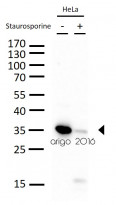
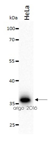
ARG20001 anti-Caspase 3 antibody [C33] WB image
Western blot: 30 µg of HeLa cells untreated or treated with Staurosporine (1μM, overnight). The blots were stained with ARG20001 anti-Caspase 3 antibody [C33] at 1:500 dilution.
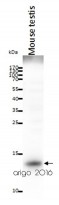
ARG20006 anti-Cytochrome C antibody WB image
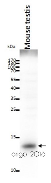
Western blot: 20 µg of Mouse testis lysate stained with ARG20006 anti-Cytochrome C antibody at 2 µg/ml dilution.
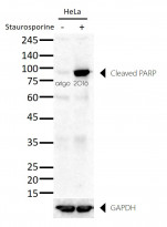
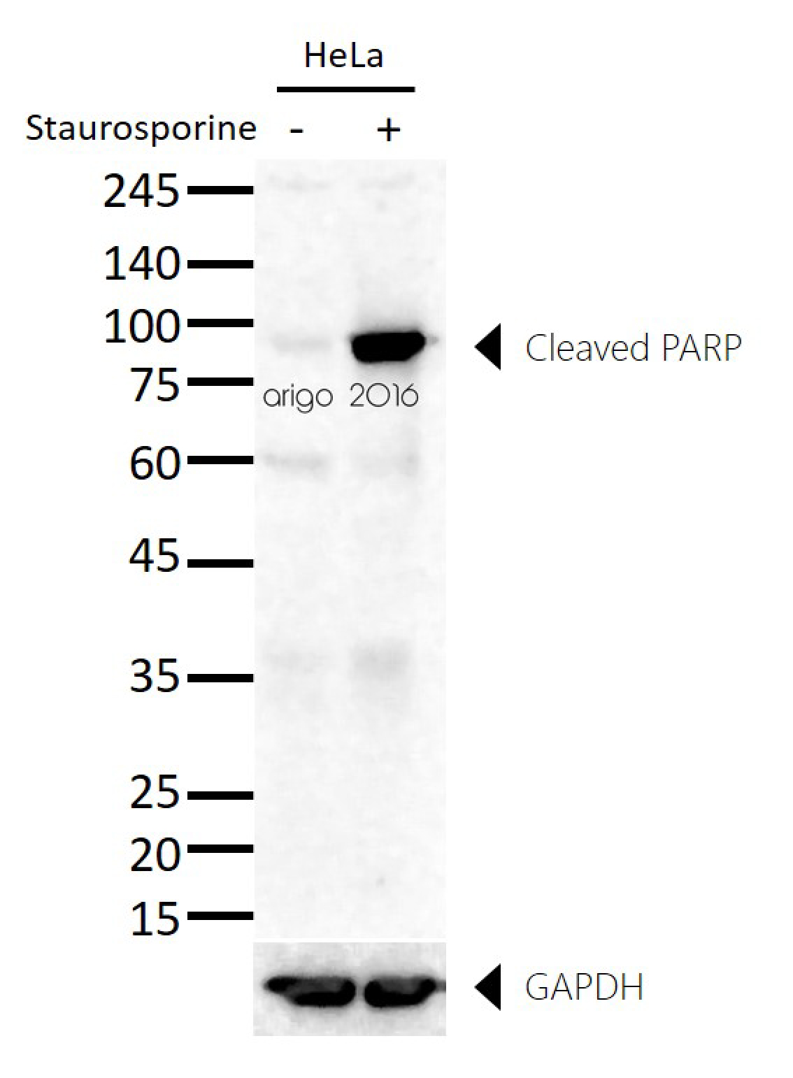
ARG20041 anti-PARP (cleaved) antibody WB image
Western blot: 30 µg of HeLa untreated or treated with Staurosporine and stained with ARG20041 anti-PARP (cleaved) antibody at 1:500 dilution.
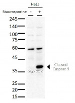
ARG20048 anti-Caspase 9 (cleaved) antibody WB image
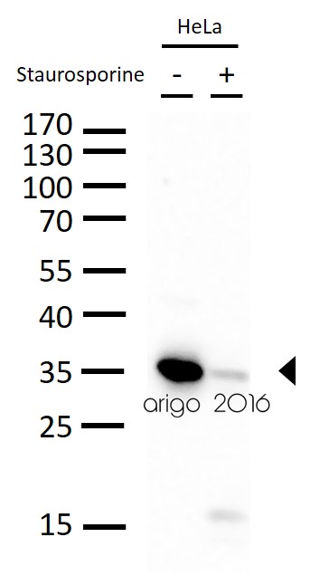
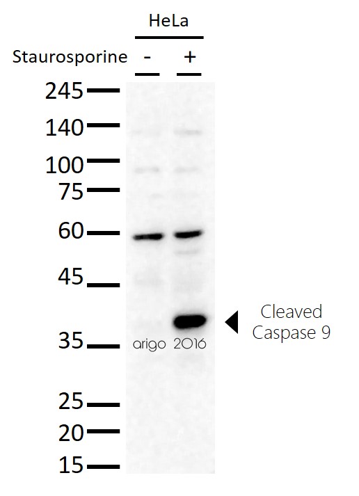
Western blot: 20 µg of HeLa untreated or treated with Staurosporine and stained with ARG20048 anti-Caspase 9 (cleaved) antibody at 1:500 dilution.
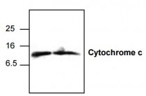
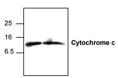
ARG20006 anti-Cytochrome c antibody WB image
Western Blot: 1. Jurkat cell lysate 2. 3T3 cell lysate stained with anti-Cytochrome c antibody (ARG20006).
The cytochrome c antibody detects the 12.6 kDa cytochrome c from human, mouse, and rat samples. Jurkat cell lysate, NIH3T3 cell lysate, and rat kidney tissue lysate can be used as positive controls.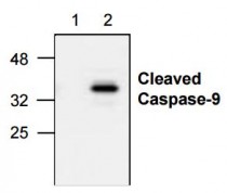
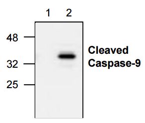
ARG20048 anti-Caspase-9 (Active) antibody WB image
Western Blot: 1. Untreated Jurkat cell lysate 2. Etoposide treated Jurkat cell lysate stained with anti-Caspase 9 active antibody (ARG20048).
The anti-active caspase-9 antibody recognizes only the cleaved caspase-9 (37 kDa). It does not recognize full-le ngth caspase-9 or any other caspases.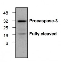
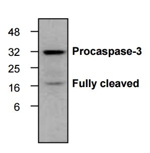
ARG20001 Caspase 3 antibody [C33] WB validated image
Western blot: HeLa cells treated with camptothecin (2 μM) stained with anti-Caspase 3 antibody [C33] (ARG20001).
The antibody recognizes both proform and the cleaved large fragment of caspase-3 in samples of human, mouse and rat origins. However, the optimal conditions should be determined individually.ARG20001 anti-Caspase 3 antibody [C33] WB image
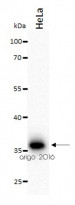
Western blot: 20 µg of HeLa cell lysates stained with ARG20001 anti-Caspase 3 antibody [C33] at 2 µg/ml dilution.


Moonlighting glyceraldehyde-3-phosphate dehydrogenase of Lactobacillus gasseri inhibits keratinocyte apoptosis and skin inflammation in experimental atopic dermatitis
ARG20001: WB / Mouse


KDF1 Promoted Proliferation, Migration and Invasion of Lung Adenocarcinoma Cells through Activating STAT3 and AKT Pathway
ARG65350: WB /
Single-Cell Analysis of Signaling Proteins Provides Insights into Proapoptotic Properties of Anticancer Drugs.
ARG20006: scPISA / Human
Probing apoptosis signaling proteins in single living cells for precision efficacy evaluation of anti-cancer drugs.
ARG20006: / Human
Inhibition of histone acetylation by curcumin reduces alcohol-induced fetal cardiac apoptosis.
ARG20001: WB / Mouse
New Products


