


MUC1 Rabbit Monoclonal Antibody(ARB993)

Key features and details
- Target:
- Clone ID:
- Host:
- Molecular Weight:
- Purity:
- Species Cross-reactivity:
- Applications:
- Swissprot ID:
-
Brand:

CAT.NO. : ARB6785
RMB Please choose
RMB Please choose
Size:
Trail, Bulk size or Custom requests Please contact us
 Product Details
Background
MUC1 (Mucin 1) is a transmembrane glycoprotein that protects epithelial cells from injury caused by external stimuli. In addition to this role, MUC1 is involved in cell-cell adhesion, proliferation, motility, invasion and survival. In epithelial cells, MUC1 expression is regulated by binding of TNFα to TNFR1 and activation of the NFκB pathway.
MUC1 is present in a variety of glandular (secretory) epithelia such as breast, eccrine and apocrine glands, and pancreas, whereas little or no MUC1 is expressed in the gastroepithelial epithelium, endocervical epithelium, and prostate glands. The immunoreactivity is usually limited to apical cell membranes, but a staining of the Golgi zone may also be seen. MUC1 can be demonstrated in most types of adenocarcinomas derived from secretory epithelia. In well-differentiated carcinomas, the staining is mostly in apical (apical) cell membranes while in the less differentiated carcinomas, a cytoplasmic staining is seen with loss of staining polarity in the membranes.
MUC1 was used to recognize epithelial differentiation when cytokeratin was difficult to visualize. In the classification of hematolymphoid neoplasms, a positive MUC1 reaction may be an aid in the identification of diffuse large B-cell lymphoma, plasma cell neoplasms and nodular lymphocyte predominant Hodgkin's lymphoma.
Application
|
Application |
Dilution Ratio |
|
IHC |
1:100 - 1:200 |
Overview
|
Predicted Molecular Wt |
122kDa |
|
Species Cross-reactivity |
Human |
|
Applications |
IHC-P |
|
Purity |
ProA affinity purified IgG |
|
Form |
Liquid |
|
Swissprot ID |
P15941 |
|
Subcellular location |
Membrane/Cytoplasm |
|
Recommended method |
Heat induced epitope retrieval with Tris-EDTA buffer (pH 9.0), primary antibody incubate at RT (18℃-25℃) for 30 minutes |
|
Immunogen |
Synthetic peptide corresponding to MUC1 residues within aa1155-1255 of MUC1 was used as an immunogen |
|
Storage Buffer |
PBS 59%, Sodium azide 0.01%, Glycerol 40%, BSA 0.05% |
Data
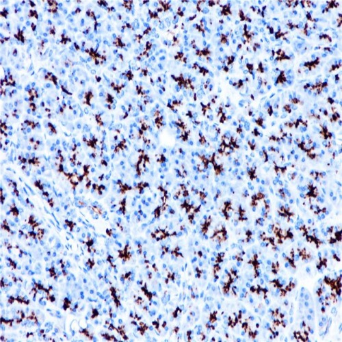
Immunohistochemical staining of human pancreas tissue using MUC1 Rabbit Monoclonal Antibody(ARB993)
Storage
Store at -20°C. Stable for one year from the date of shipment.
Research Use Only
For Research Use Only. Not for use in diagnostic procedures.
New Products


