


Neurite Marker Antibody Duo


Key features and details
- 产品描述:
- 靶点名称:
- 別名:
-
Brand:

 Product Details
| 货号 | 内含物名称 | 宿主克隆性 | 反应 | 应用 | 包装 |
|---|---|---|---|---|---|
| ARG52328 | anti-MAP2 antibody | Chicken pAb | Hu, Ms, Rat, Bov, Dog, Marmoset, Sheep | ICC/IF, IHC-FoFr , IHC-Fr, IHC-P, WB | 50 μl |
| ARG62683 | anti-beta III Tubulin antibody [TU-20] | Mouse mAb | Hu, Ms, Rat, Dog, Pig | FACS, ICC/IF, IHC-Fr, IHC-P, WB | 50 μg |
| 产品描述 | Neurites refer to projections from the soma of a neuron. The projection can be either an axon or a dendrite. Axons and dendrites are molecularly and functionally distinct, the slender axon is required for information transmission to other cells, whereas the branched dendrites are responsible for receiving electrochemical stimuli from other neurons. Establishment of the axon/dendrite polarity is critical for the neuronal functions. beta III Tubulin antibody is a well-known axonal marker and MAP2 antibody is excellent for staining dendrites.arigo's Neurite Marker Antibody Duo comprises beta III Tubulin and MAP2 antibodies and is an excellent for studying neuronal differentiation. This antibody Duo can be also used as a neuron marker duo for labeling neuronal cells. Related news: Astrocyte-to-neuron conversion for Parkinson's disease treatment |
|---|---|
| 靶点名称 | Neurite Marker |
| 別名 | Neurite Marker antibody; MAP2 antibody; beta III Tubulin antibody |
| 存放说明 | For continuous use, store undiluted antibody at 2-8°C for up to a week. For long-term storage, aliquot and store at -20°C or below. Storage in frost free freezers is not recommended. Avoid repeated freeze/thaw cycles. Suggest spin the vial prior to opening. The antibody solution should be gently mixed before use. |
|---|---|
| 注意事项 | For laboratory research only, not for drug, diagnostic or other use. |
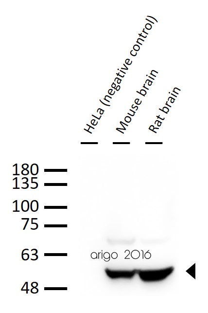
ARG62683 anti-beta III Tubulin antibody [TU-20] WB image
Western blot: 30 µg of 1) HeLa (negative control), 2) Mouse brain, and 3) Rat brain lysate stained with ARG62683 anti-beta III Tubulin antibody [TU-20] at 1:1000 dilution.
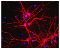
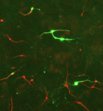
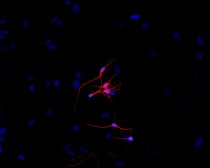
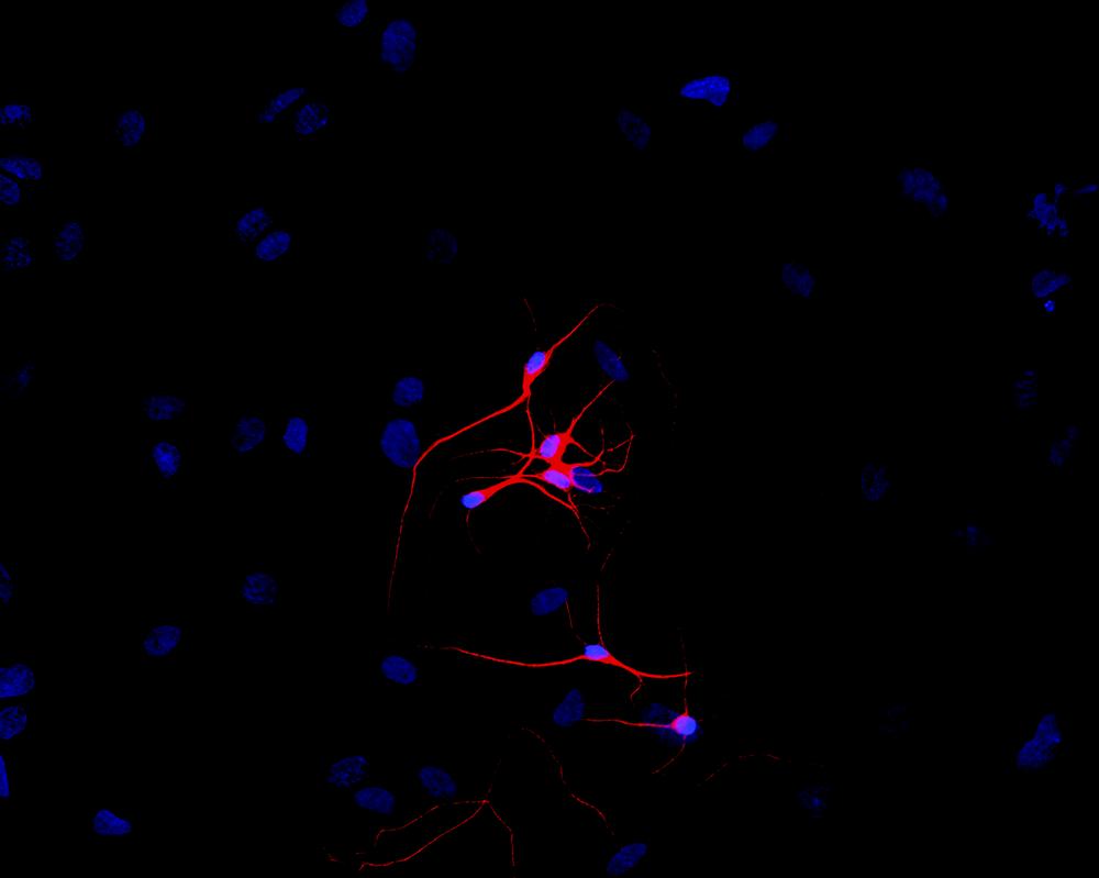
ARG52328 anti-MAP2 antibody ICC/IF image
Immunofluorescence: Mixed neuron/glial cultures. The perikarya and dendrites of neurons are strongly and specifically stained with ARG52328 anti-MAP2 antibody (red). Cell nuclei are visualized with DAPI DNA stain.
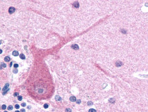
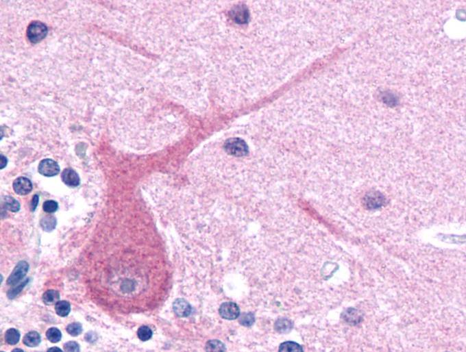
ARG62683 anti-beta III Tubulin antibody [TU-20] IHC-P image
Immunohistochemistry: Paraffin-embedded Human brain tissue stained with ARG62683 anti-beta III Tubulin antibody [TU-20].
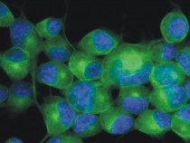
ARG62683 anti-beta III Tubulin antibody [TU-20] ICC/IF image
Immunofluorescence: Neuro2a mouse neuroblastoma cell stained with ARG62683 anti-beta III Tubulin antibody [TU-20] (green).
Cell nuclei was stained with DAPI (blue).
ARG52328 anti-MAP2 antibody WB image
Western blot: Rat cortex lysate stained with ARG52328 anti-MAP2 antibody showing specific immunolabeling of the ~ 280 kDa MAP2 protein.
ARG62683 anti-beta III Tubulin antibody [TU-20] IHC-P image
Immunohistochemistry: Mouse brain stained with ARG62683 anti-beta III Tubulin antibody [TU-20].
ARG62683 anti-beta III Tubulin antibody [TU-20] ICC/IF image
Immunofluorescence: P-19 mouse embryonal carcinoma cells stimulated to neuronal differentiation by retinoic acid stained with ARG62683 anti-beta III Tubulin antibody [TU-20] (red).
Cell nuclei was stained with DAPI (blue).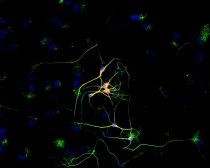
ARG62683 anti-beta III Tubulin antibody [TU-20] ICC/IF image
Immunofluorescence: P-19 mouse embryonal carcinoma cell line stimulated to neuronal differentiation by retinoic acid co-stained with stained with ARG62683 anti-beta III Tubulin antibody [TU-20] (red) and anti-beta-tubulin (green)
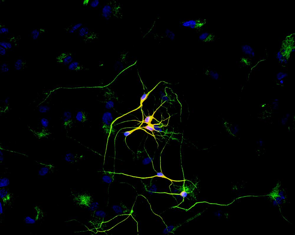
Superposition of red and green colours provided yellow staining. Nuclei were stained with DNA-binding dye (blue).ARG52328 anti-MAP2 antibody ICC/IF image
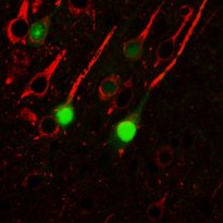
Immunofluorescence: E17 Rat midbrain mixed neuronal cultures stained with ARG52461 anti-Tyrosine Hydroxylase antibody (green) and ARG52328 anti-MAP2 antibody (red).


Magnetic stirring with iron oxide nanospinners accretes neurotoxic Aβ42 oligomers into phagocytic clearable plaques for Alzheimer's disease treatment
ARG52328: ICC/IF / Mouse


Extracellular RNAs-TLR3 signaling contributes to cognitive decline in a mouse model of postoperative cognitive dysfunction.
ARG52328: IHC-Fr / Mouse
New Products


