


Osteocalcin Rabbit Polyclonal Antibody


Key features and details
- Reactivity:Human, Mouse, Rat
- Application:WB, IHC, IF/ICC
- Host:Rabbit
- Clonality:Polyclonal
- Target:Osteocalcin
-
Brand:

CAT.NO. : ARA6678
RMB Please choose
RMB Please choose
*产品价格可能会有所调整,请以品牌方官网实时更新的价格为准,以确保准确性。
 Product Details
Background
Osteocalcin belongs to the osteocalcin/matrix Gla-protein family and constitutes 1-2% of the total bone protein. It binds strongly to apatite and calcium. Gamma-carboxyglutamate residues are formed by vitamin K dependent carboxylation. These residues are essential for the binding of calcium.
Application
|
Application |
Dilution Ratio |
|
WB |
1:500 - 1:1000 |
|
IHC |
1:50 - 1:200 |
|
IF/ICC |
1:50 - 1:200 |
Overview
|
Immunogen |
KLH-conjugated synthetic peptide encompassing a sequence within the center region of human Osteocalcin. The exact sequence is proprietary |
|
Purification Method |
The antibody was purified by immunogen affinity chromatography |
|
Clonality |
Polyclonal |
|
Form |
Liquid in 0.42% Potassium phosphate, 0.87% Sodium chloride, pH 7.3, 30% glycerol, and 0.01% sodium azide |
|
Gene Symbol |
BGLAP |
|
Alternative Names |
Osteocalcin; Bone Gla protein; BGP; Gamma-carboxyglutamic acid-containing protein |
|
Gene ID (Human) |
632 |
|
Gene ID (Mouse) |
12096 |
|
Gene ID (Rat) |
25295 |
|
Protein ID (Human) |
P02818 |
|
Protein ID (Mouse) |
P86546 |
|
Protein ID (Rat) |
P04640 |
Data
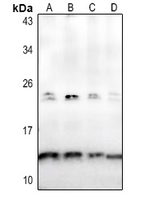
Western blot analysis of Osteocalcin expression in HCT116 (A), HEK293T (B), PC12 (C), NIH3T3 (D) whole cell lysates. (Predicted band size: 10 kD; Observed band size: 13 kD)
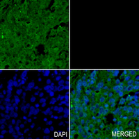
Immunohistochemical analysis of Osteocalcin staining in human lung cancer formalin fixed paraffin embedded tissue section. The section was pre-treated using heat mediated antigen retrieval with sodium citrate buffer (pH 6.0). The section was then incubated with the antibody at room temperature and detected using an HRP conjugated compact polymer system. Tyramide-AF488 (green) was used as the chromogen. The section was then counterstained with DAPI (blue).
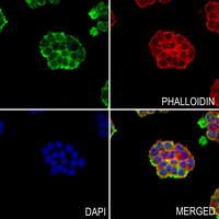
Immunofluorescent analysis of Osteocalcin staining in MDAMB231 cells. Formalin-fixed cells were permeabilized with 0.1% Triton X-100 in TBS for 5-10 minutes and blocked with 3% BSA-PBS for 30 minutes at room temperature. Cells were probed with the primary antibody in 3% BSA-PBS and incubated overnight at 4 °C in a hidified chamber. Cells were washed with PBST and incubated with a AF488-conjugated secondary antibody (green) in PBS at room temperature in the dark. Phalloidin - AF594 was used to stain Actin filaments (red). DAPI was used to stain the cell nuclei (blue).
Storage
Store at 4°C short term. For long term storage, store at -20°C, avoiding freeze/thaw cycles.
Research Use Only
For Research Use Only. Not for use in diagnostic procedures.
New Products


