


Phospho-Erk1 (T202 + Y204) + Erk2 (T185 + Y187) Rabbit Monoclonal Antibody(ARA723)
Key features and details
- Reactivity:Human, Mouse, Rat
- Application:WB, IHC, IF/ICC, FC
- Host:Rabbit
- Clonality:Monoclonal
- lsotype: IgG
- Target:Phospho-Erk1 (T202 + Y204) + Erk2 (T185 + Y187)
-
Brand:

 Product Details
|
Application |
Dilution Ratio |
|
WB |
1:5,000 - 1:10,000 |
|
IF/ICC |
1:80 - 1:100 |
|
IHC |
1:800 - 1:1,000 |
|
FC |
1:800 - 1:1,000 |
|
Antibody Type |
Recombinant Rabbit monoclonal Antibody |
|
Immunogen |
Synthetic peptide within Human ERK1 aa 166-215 / 379. |
|
Species Reactivity |
Human, Mouse, Rat |
|
Validated Applications |
WB, IHC, IF/ICC, FC |
|
Molecular Weight |
Predicted band size: 41/43 kDa |
|
Positive Control |
SH-SY5Y cell lysate, SH-SY5Y treated with 100ng/mL hβ-NGF for 10 minutes cell lysate, PC-12 treated with 100ng/mL hβ-NGF for 10 minutes cell lysate, SK-Br-3 whole cell lysate, NIH/3T3 cell lysate, NIH/3T3 treated with 200nM PMA for 30 minutes cell lysate, HeLa treated with 200nM PMA for 30 minutes cell lysate, human thyroid carcinoma tissue. |
|
Conjugation |
unconjugated |
|
Form |
Liquid |
|
Storage Buffer |
1×TBS (pH7.4), 0.05% BSA, 40% Glycerol. Preservative: 0.05% Sodium Azide. |
|
Isotype |
IgG |
|
Purification Method |
Protein A affinity purified. |

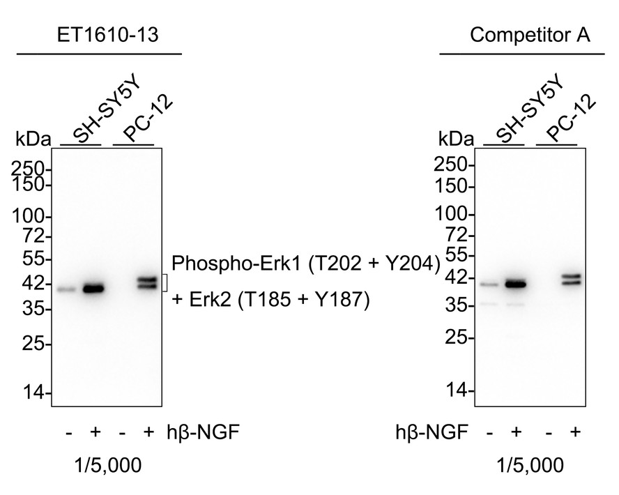
Western blot analysis of Phospho-Erk1 (T202 + Y204) + Erk2 (T185 + Y187) on different lysates with Phospho-Erk1 (T202 + Y204) + Erk2 (T185 + Y187) Rabbit Monoclonal Antibody(ARA723).
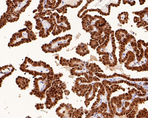
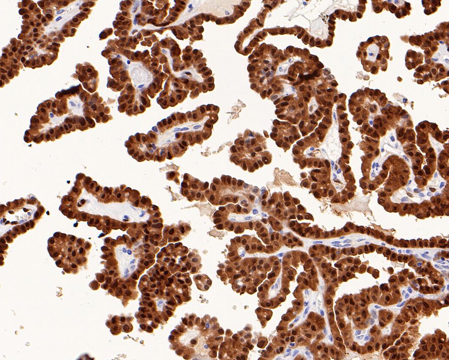
Immunohistochemical analysis of paraffin-embedded human thyroid carcinoma tissue with Phospho-Erk1 (T202 + Y204) + Erk2 (T185 + Y187) Rabbit Monoclonal Antibody(ARA723).

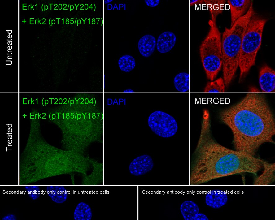
Immunocytochemistry analysis of NIH/3T3 cells treated with 200nM PMA for 30 minutes labeling Phospho-Erk1 (T202 + Y204) + Erk2 (T185 + Y187) with Phospho-Erk1 (T202 + Y204) + Erk2 (T185 + Y187) Rabbit Monoclonal Antibody(ARA723).
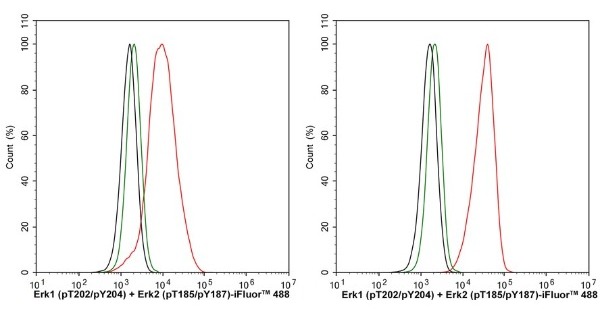
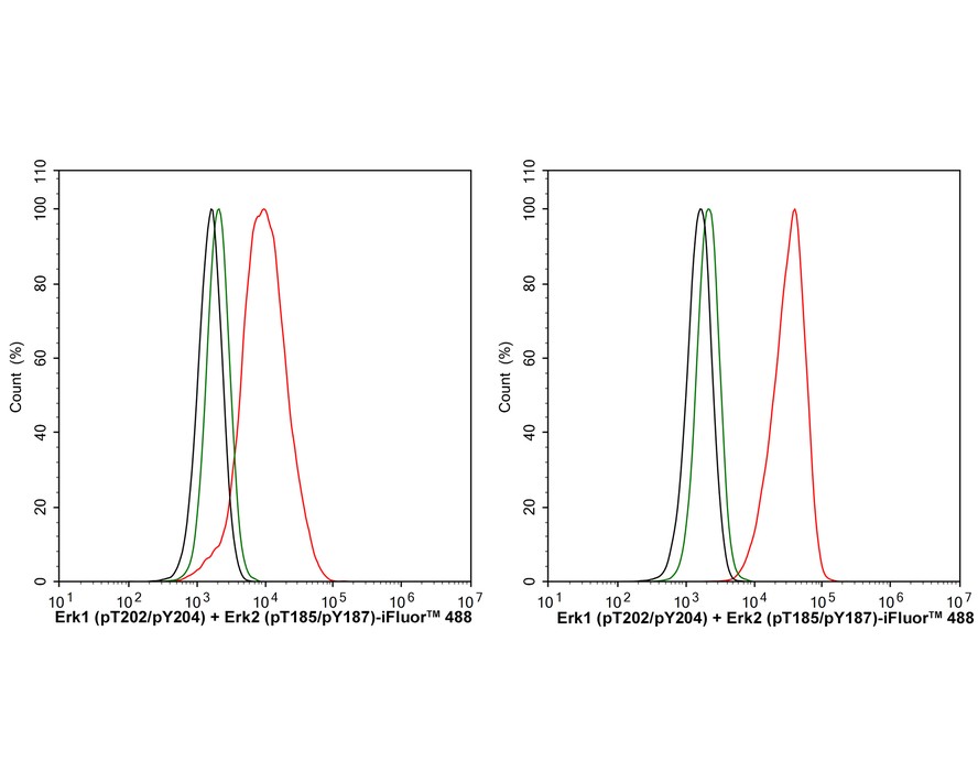
Flow cytometric analysis of HeLa cells untreated (left) or treated (right) with PMA for 30 minutes labeling Phospho-Erk1 (T202 + Y204) + Erk2 (T185 + Y187).
New Products


