


SDHA Rabbit Polyclonal Antibody

Key features and details
- Reactivity:Human, Mouse, Rat
- Application:WB,IHC, IF/ICC
- Host:Rabbit
- Clonality:Polyclonal
- Target:SDHA
-
Brand:

CAT.NO. : ARA6635
RMB Please choose
RMB Please choose
Size:
Trail, Bulk size or Custom requests Please contact us
*产品价格可能会有所调整,请以品牌方官网实时更新的价格为准,以确保准确性。
 Product Details
Background
This gene encodes a major catalytic subunit of succinate-ubiquinone oxidoreductase, a complex of the mitochondrial respiratory chain. The complex is composed of four nuclear-encoded subunits and is localized in the mitochondrial inner membrane. Mutations in this gene have been associated with a form of mitochondrial respiratory chain deficiency known as Leigh Syndrome. A pseudogene has been identified on chromosome 3q29.
Application
| Application | Dilution Ratio |
|---|---|
| WB | 1:500 - 1:2000 |
| IHC | 1:50 - 1:200 |
| IF/ICC | 1:50 - 1:200 |
Overview
|
Product Description |
Rabbit polyclonal antibody to SDHA |
|
Immunogen |
Recombinant full length protein of human SDHA |
|
Purification Method |
The antibody was purified by immunogen affinity chromatography |
|
Clonality |
Polyclonal |
|
Product Form |
Liquid in 0.42% Potassium phosphate, 0.87% Sodium chloride, pH 7.3, 30% glycerol, and 0.01% sodium azide |
|
Gene Name |
SDHA |
|
Related Names |
SDH2; SDHF; Succinate dehydrogenase [ubiquinone] flavoprotein subunit mitochondrial; Flavoprotein subunit of complex II |
|
Gene ID (Human) |
6389 |
|
Gene ID (Mouse) |
66945 |
|
Gene ID (Rat) |
157074 |
|
Protein ID (Human) |
P31040 |
|
Protein ID (Mouse) |
Q8K2B3 |
|
Protein ID (Rat) |
Q920L2 |
Data

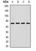
Western blot analysis of SDHA expression in HepG2 (A), MCF7 (B), mouse liver (C), mouse stomach (D) whole cell lysates. (Predicted band size: 56; 67; 72 kD; Observed band size: 70 kD)
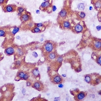
Immunohistochemical analysis of SDHA staining in human liver cancer formalin fixed paraffin embedded tissue section. The section was pre-treated using heat mediated antigen retrieval with sodium citrate buffer (pH 6.0). The section was then incubated with the antibody at room temperature and detected using an HRP conjugated compact polymer system. DAB was used as the chromogen. The section was then counterstained with haematoxylin and mounted with DPX.
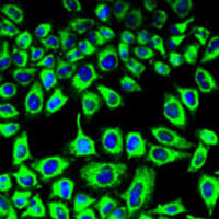
Immunofluorescent analysis of SDHA staining in HeLa cells. Formalin-fixed cells were permeabilized with 0.1% Triton X-100 in TBS for 5-10 minutes and blocked with 3% BSA-PBS for 30 minutes at room temperature. Cells were probed with the primary antibody in 3% BSA-PBS and incubated overnight at 4 °C in a humidified chamber. Cells were washed with PBST and incubated with a AF488-conjugated secondary antibody (green) in PBS at room temperature in the dark. DAPI was used to stain the cell nuclei (blue).
Storage
Store at 4°C short term. For long term storage, store at -20°C, avoiding freeze/thaw cycles.
Research Use Only
For Research Use Only. Not for use in diagnostic procedures.
New Products


