


Tau (S305) Mouse Monoclonal Antibody(ARA971)

Key features and details
- Reactivity:Human, Rat, Mouse
- Application:WB, ELISA, IHC-P, IF
- Host:Mouse
- Clonality:Monoclonal
- Target:Tau (S305)
-
Brand:

CAT.NO. : ARA6837
RMB Please choose
RMB Please choose
*产品价格可能会有所调整,请以品牌方官网实时更新的价格为准,以确保准确性。
 Product Details
Background
Tau (S305) refers to a specific site on the tau protein, where a mutation in the S305N form is associated with a form of frontotemporal dementia. Research shows that phosphorylation at the S305 site normally inhibits tau aggregation, a process linked to neurodegenerative diseases like Alzheimer's and frontotemporal dementia. Mutations at this site, such as S305N, increase the proportion of a specific tau isoform (4R) and can disrupt normal brain cell function.
Application
|
Application |
Dilution Ratio |
|
WB |
1:800 - 1:1000 |
|
Sandwich ELISA |
1:250 - 1:500 |
|
IHC-P |
1:150 - 1:200 |
|
IF |
1:200 - 1:250 |
Overview
|
Antibody Specificity |
Recognizing beta Amyloid 1-40 with an epitope within 15 amino acids at the C-terminus of beta Amyloid 1-40 |
|
Species Reactivity |
Human |
|
Clonality |
Monoclonal |
|
Host / Isotype |
Mouse / IgG1 |
|
Immunogen |
Human amyloid beta precursor peptide |
|
Cross Reactivity |
Not tested |
|
Recombinant |
Not recombinant |
|
Purification |
Protein A purification |
|
Form |
Liquid |
|
Conjugation |
Unconjugated |
Data
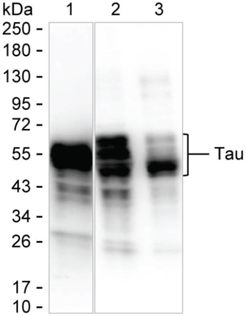
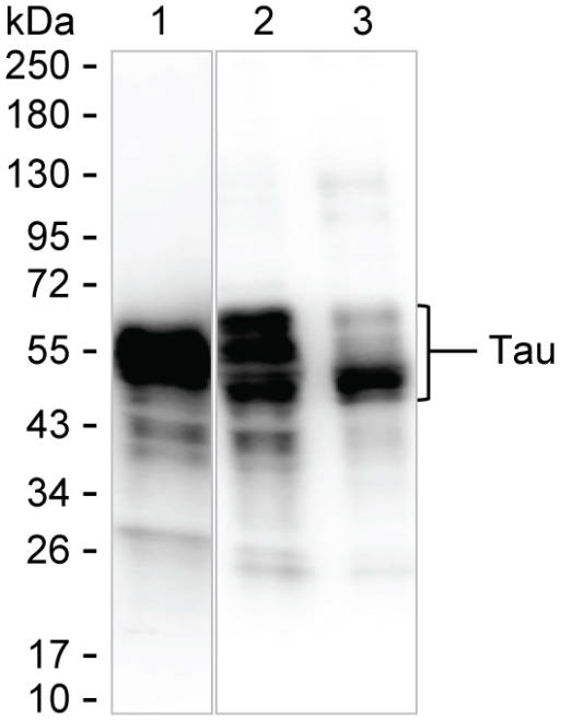
Western blotting analysis of Tau (S305) by ARA6837.
Various protein samples were run on 6-18% SDS-PAGE under reducing conditions and blotted onto nitrocellulose membrane. ARA6837 was used as the primary antibody and peroxidase conjugated goat anti-mouse IgG was used as the secondary antibody. Tau band was visualized using ECL Western Blotting Substrate.
Lane 1: 15 μg of brain tissue lysate
Lane 2: 15 μg of rat brain tissue lysate
Lane 3: 15 μg of mouse brain tissue2 lysate
Result: ARA6837 can detect Tau by Western blotting.

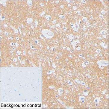
IHC-P analysis of brain tissue by Tau antibody (ARA6837).
IHC-P was performed using sections of formalin-fixed, paraffin-embedded brain tissue. Antigen was retrieved through addition of boiling Tris/EDTA buffer pH 9 in a pressure cooker for 3 min. Endogenous peroxidase activity was quenched by incubating the sections with 3% H₂O₂ for 30 min at room temperature. The sections were then incubated with primary antibody (ARA6837) at room temperature for 1 h. Poly-peroxidase conjugated goat anti-mouse IgG was used as the secondary antibody. Diaminobenzidine was used as the chromogen. The section was counterstained with hematoxylin. A tissue section incubated with phosphate-buffered saline followed by incubation with the secondary antibody was used as the background control.
Result: Neuropil is positively stained.
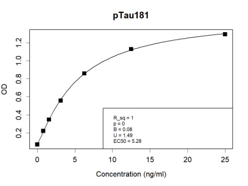
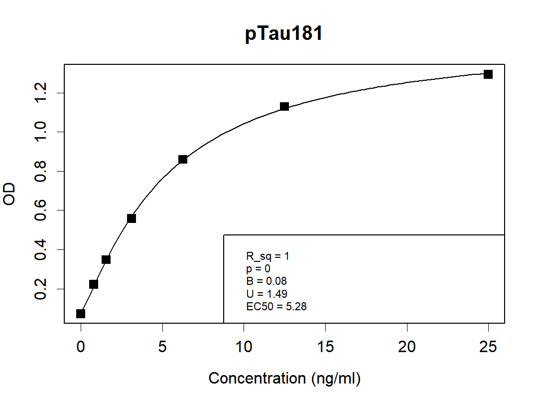
Sandwich ELISA analysis of Tau (S305) by ARA6837 & ARA6844.
Microtiter plates were coated with ARA6844 as the capture antibody. Recombinant pTau181 protein (Cat. AXP6613) was applied as the antigen.Peroxidase conjugated pTau181 Antibody (ARA6837) served as the detection antibody.
Result: ARA6837 and ARA6844 can be used as a matched antibody pair to detect and quantify the concentration of pTau181.
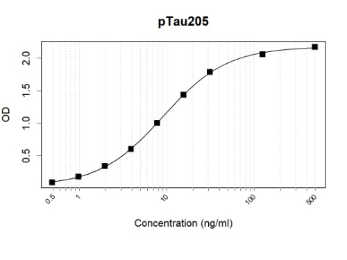
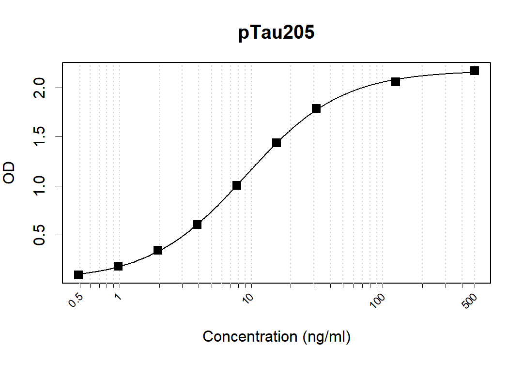
Sandwich ELISA analysis of Tau (S305) by ARA6837 & ARA6848.
Microtiter plates were coated with ARA6848 as the capture antibody. Recombinant pTau205 protein (Cat. AXP6615) was applied as the antigen.Biotin-conjugated pTau205 Antibody (ARA6837) served as the detection antibody, followed by streptavidin-HRP incubation.
Result: ARA6837 and ARA6848 can be used as a matched antibody pair to detect and quantify the concentration of pTau205.

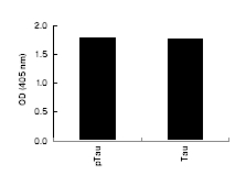
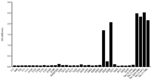
Cross reactivity of ARA6837 to other peptides and recombinant proteins by ELISA. Microtiter wells were coated with various peptides and recombinant proteins.
ARA6837 was used as the primary antibody and peroxidase conjugated goat anti-mouse IgG was used as the secondary antibody.
Result: ARA6837 can detect Tau proteins regardless of phosphorylation.

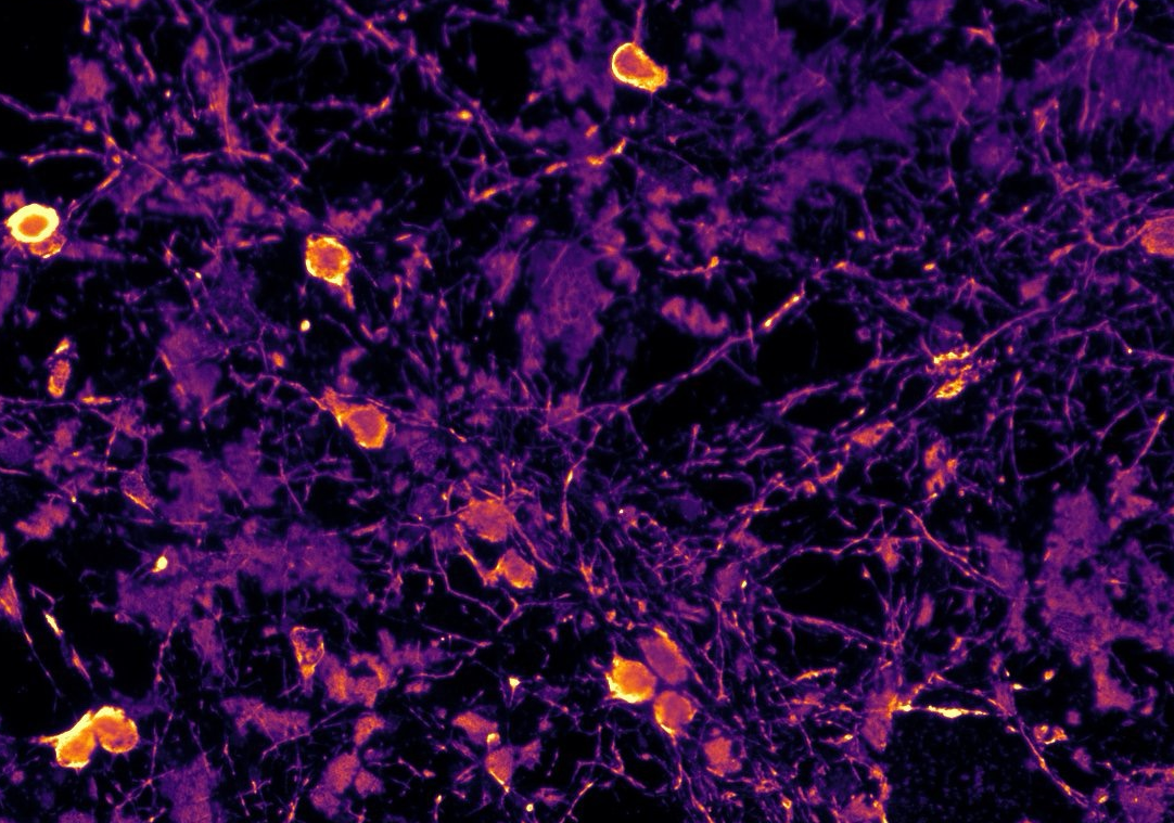
Immunofluorescence analysis of Tau (S305) by ARA6837. Immunofluorescence analysis of primary rat neurons by Tau (S305) antibody (ARA6837). The cells were fixed, permeabilized, blocked and incubated with the primary antibody (1:250). Followed by secondary antibody staining, nuclei were counterstained.
Epitope analysis of Tau (S305) by ARA6837 by ELISA.
Synthetic peptides containing indicated residues were coated onto microtiter wells. Recombinant Tau (2-244), Tau (1-368), Tau (243-368) and Tau (1-441) proteins with isoform P10636-8 or Tau (1-758) with isoform P10636-1 were also coated. The antibody ARA6837 was applied as the primary antibody. Peroxidase-conjugated goat anti-mouse IgG was used as the secondary antibody, and signals were detected using a colorimetric assay.
Result: ARA6837 recognizes Tau protein containing S305 and S356 peptides that share a sequence of HVPGGG.
Storage
Store at +4℃ after thawing. Aliquot store at -20℃. Avoid repeated freeze / thaw cycles.
Research Use Only
For Research Use Only. Not for use in diagnostic procedures.
New Products


