


WT1 Rabbit Monoclonal Antibody(ARB572)
Key features and details
- Target:
- Clone ID:
- Host:
- Molecular Weight:
- Purity:
- Species Cross-reactivity:
- Applications:
- Swissprot ID:
-
Brand:

CAT.NO. : ARB6864
RMB Please choose
RMB Please choose
Size:
Trail, Bulk size or Custom requests Please contact us
 Product Details
Background
The WT1 gene located at chromosome 11p13 codes for a transcription factor, a DNA - binding nucleoprotein, that plays a role primarily in the development of genitourinary organs. There are at least eight isoforms ranging between 52 and 62 kDa produced by combination of alternative splicing and RNA editing. WT1 is synthesized and reside in the cytoplasm in an inactive form. When activated through phosphorylation it is translocated to the nucleus. WT1 influences cell proliferation by suppressing bcl - 2 and regulating cadherin and p53.
In normal epithelia, nuclear WT1 expression is largely restricted to ovary (surface epithelium and inclusion cysts) and fallopian tube, while WT1 is not found in endometrial or cervical epithelium. As regards nonepithelial cells, nuclear WT1 is found in mesothelium and some submesothelial stromal cells, stromal cells of the female genital tract, testicular non - germinal cells, and kidney (podocytes). In tumor tissues, WT1 is detected in tumor cells of Wilms’ Tumor (also known as nephroblastoma) and mesothelioma. Additionally, WT1 expression has been found in ovarian serous carcinomas and some breast carcinomas. WT1 is particularly used for distinguishing malignant mesothelioma and ovarian serous carcinoma from nonserous carcinomas. As for malignant mesothelioma, calretinin and WT1 are superior to cytokeratin 5/6, N - cadherin and thrombomodulin. WT1 is also applicable for the differential diagnostic of small childhood tumors.
In normal epithelia, nuclear WT1 expression is largely restricted to ovary (surface epithelium and inclusion cysts) and fallopian tube, while WT1 is not found in endometrial or cervical epithelium. As regards nonepithelial cells, nuclear WT1 is found in mesothelium and some submesothelial stromal cells, stromal cells of the female genital tract, testicular non - germinal cells, and kidney (podocytes). In tumor tissues, WT1 is detected in tumor cells of Wilms’ Tumor (also known as nephroblastoma) and mesothelioma. Additionally, WT1 expression has been found in ovarian serous carcinomas and some breast carcinomas. WT1 is particularly used for distinguishing malignant mesothelioma and ovarian serous carcinoma from nonserous carcinomas. As for malignant mesothelioma, calretinin and WT1 are superior to cytokeratin 5/6, N - cadherin and thrombomodulin. WT1 is also applicable for the differential diagnostic of small childhood tumors.
Application
|
Application |
Dilution Ratio |
|
IHC |
1:100 - 1:200 |
Overview
|
Predicted Molecular Wt |
49kDa |
|
Species Cross-reactivity |
Human |
|
Applications |
IHC-P |
|
Purity |
ProA affinity purified IgG |
|
Form |
Liquid |
|
Swissprot ID |
P19544 |
|
Subcellular location |
Nucleus |
|
Recommended method |
Heat induced epitope retrieval with Tris-EDTA buffer (pH 9.0), primary antibody incubate at RT (18℃-25℃) for 30 minutes |
|
Immunogen |
Synthetic peptide corresponding to WT1 residues within aa1 - 100 of WT1 was used as an immunogen |
|
Storage Buffer |
PBS 59%, Sodium azide 0.01%, Glycerol 40%, BSA 0.05% |
Data

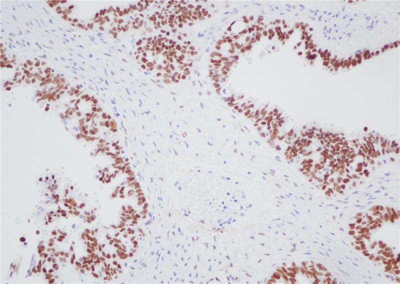
Immunohistochemical staining of human ovarian cancer tissue using WT1 Rabbit Monoclonal Antibody(ARB572)
Storage
Store at -20°C. Stable for one year from the date of shipment.
Research Use Only
For Research Use Only. Not for use in diagnostic procedures.
New Products


