


ZO1 Rabbit Polyclonal Antibody
Key features and details
- Reactivity:Human, Mouse
- Application:WB, ICC/IF
- Host:Rabbit
- Clonality:polyclonal
- Target:ZO1
-
Brand:

CAT.NO. : ARA6431
RMB Please choose
RMB Please choose
*产品价格可能会有所调整,请以品牌方官网实时更新的价格为准,以确保准确性。
 Product Details
Background
ZO-1 forms complexes with either ZO-2 or ZO-3. In addition, these proteins can also associate with claudin, occludin and F-actin, at tight junction stands, where they provide a linkage between the actin cytoskeleton and the tight junction. ZO-1 expression is significantly reduced in many breast cancer lines. ZO-2 and ZO-3 are ubiquitously expressed within epithelial tight junctions, and unlike ZO-1, which is also expressed at cell junctions of cardiac myocytes, ZO-2 is not expressed in nonepithelial tissue. ZO-1 is a protein located on a cytoplasmic membrane surface of intercellular tight junctions. ZO-1 may be involved in signal transduction at cell-cell junctions. Two transcript variants encoding distinct isoforms have been identified for the ZO-1 gene. Diseases associated with ZO-1 dysfunction include Celiac Disease and Congenital Nephrotic Syndrome.
Application
|
Application |
Dilution Ratio |
|
WB |
1:500 - 1:1000 |
|
ICC/IF |
1:50 - 1:200 |
Overview
|
Product Description |
Rabbit polyclonal antibody to ZO1 |
|
Immunogen |
KLH-conjugated synthetic peptide of human ZO1 |
|
Purification Method |
The antibody was purified by immunogen affinity chromatography. |
|
Clonality |
Polyclonal |
|
Form |
Liquid in 0.42% Potassium phosphate, 0.87% Sodium chloride, pH 7.3, 30% glycerol, and 0.01% sodium azide. |
|
Gene Symbol |
TJP1 |
|
Alternative Names |
ZO1; Tight junction protein ZO-1; Tight junction protein 1; Zona occludens protein 1; Zonula occludens protein 1 |
|
Gene ID (Human) |
7082 |
|
Protein ID (Human) |
Q07157 |
Data

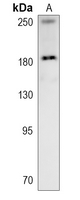
Western blot analysis of ZO1 expression in mouse colon (A) whole cell lysates. (Predicted band size: 186; 195 kD; Observed band size: 186; 240 kD)

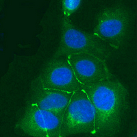
Immunofluorescent analysis of ZO1 staining in MCF7 cells. Formalin-fixed cells were permeabilized with 0.1% Triton X-100 in TBS for 5-10 minutes and blocked with 3% BSA-PBS for 30 minutes at room temperature. Cells were probed with the primary antibody in 3% BSA-PBS and incubated overnight at 4 °C in a humidified chamber. Cells were washed with PBST and incubated with a AF488-conjugated secondary antibody (green) in PBS at room temperature in the dark. DAPI was used to stain the cell nuclei (blue).
Storage
Store at 4°C short term. For long term storage, store at -20°C, avoiding freeze/thaw cycles.
Research Use Only
For Research Use Only. Not for use in diagnostic procedures.
New Products


